Rules
Criteria for Judgment
The Award will be given to the most striking image that clearly conveys a point of important biological information in Drosophila research. Selection criteria include, but are not limited to: aesthetic appeal, importance of the result, and the clarity with which it is shown. As of 2011, an Award will be given each year in both the Still Image and Video categories.
Eligibility
Because the Award is tied closely to science as well as aesthetics, only images that have been published in a primary research journal in a given calendar year are eligible for the award. The Award will be presented to the individual researcher who generated and documented the data, at whatever professional level (undergraduate, graduate student, postdoc, technician, PI); the Award will only be given to individuals. Submissions are to be made either by the researcher or by nominators with the researcher’s permission. For other eligibility questions, see the FAQ.
Guidelines for image submission
Submissions will take the Format of a two page PowerPoint presentation, of file size not to exceed 2 MB. The first page should contain a single image, which may consist of several panels, although simplicity of presentation should be kept in mind. The image must be essentially as published. For instance, rearranging and combining multiple panels is allowed; altering color is not.
Video submissions should include a .mov or .avi file of preferably 50 MB or less, accompanied by a caption and reference displayed in a .ppt file.
The second page should contain both a Title and an accompanying Caption, as complete but brief as possible, in the style of documentation for the cover of a general interest journal. Both descriptive detail and biological significance should be made clear. The caption should conclude with a Reference to the published work, and include an html link to the article on the journal site.
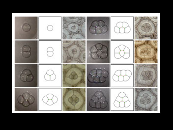
First Slide Example:

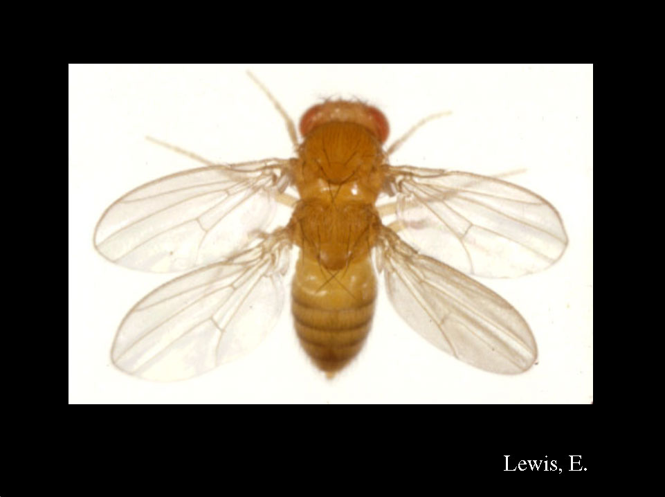
Second Slide Example:
Surface mechanics constrain cells to adopt physically stable configurations by minimizing the surface free energy. Under this restrictions, retinal cone cell clusters self-organized themselves into bubble-like patterns. The figure shows the stable patterns of soap bubble aggregates (left panels) and rough eye (Roi) mutant ommatidia (right panels), where variable numbers of cone cells develop. The extensive correlation suggests that surface minimization is essential for cone cell patterning.
Takashi Hayashi and Richard W. Carthew.
Surface mechanics mediate pattern formation in the developing retina.
Nature 431, 647-652 (2004).
Submitting an entry
Submissions are to be emailed to the following Contact Address: drosophilaimageaward@gmail.com
To submit large entries, email with the subject heading ‘Request invitation to submit Image Award via fileshare’; you will receive a link to a filesharing site for upload.
Award Process
The Image Award committee will accept electronic submissions from October to January for work published in a given calendar year. The committee will select ten finalists, who will be notified to the Drosophila Research Conference. All finalists will also have their images displayed during the presentation. The committee will select one winner from these finalists, who will be presented with a plaque and framed copy of the winning image in a plenary session presentation.
Frequently Asked Questions
Please view our FAQ’s for answers to the most common questions.

