2007 Drosophila Image Award
Winner
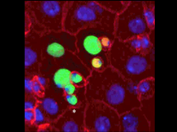
The adult Drosophila midgut, like the vertebrate intestine, is made up of enterocytes (digestive cells) and hormone producing enteroendocrine cells. Previously stem cells had not been described in flies and Drosophila intestinal cells had thought to be relatively stable.
A eight-cell intestinal stem cell clone made up of a single stem cell (asterisk), recent stem cell daughter (adjacent to the stem cell), two enteroendocrine cells (arrowheads), and one early enterocyte and three mature enterocytes (larger nuclei). Clone (green), cell membranes (red), enteroendocrine cells (nuclear, red), nucleus (blue).
This clone and ones like it , demonstrate for the first time the existence of multipotent intestinal stem cells in Drosophila. The ability to easily identify stem cells and to manipulate them genetically will facilitate the analysis of normal and abnormal intestinal function in both Drosophila and vertebrates .
Benjamin Ohlstein and Allan Spradling.
The adult Drosophila posterior midgut is maintained by pluripotent stem cells.
Nature, vol, 439, 470-473 (2006).
Runner-up
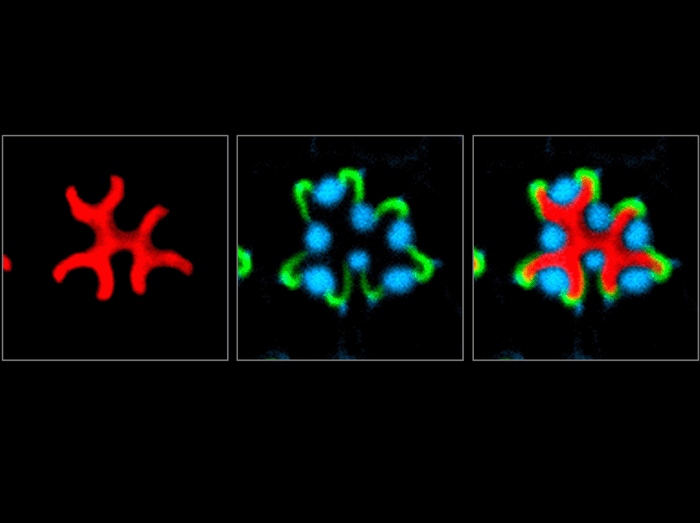
In the fly retina an epithelial lumen, called the interrhabdomeral space, forms between the photoreceptor cells. The interrhabdomeral space has a critical function in vision as it optically isolates individual photoreceptor cells. These confocal images of an adult ommatidium show that the secretion of the proteoglycan Eyes shut (red) is essential for opening up a lumen between the photoreceptor cells. Eyes shut (red) is localized exclusively to the interrhabdomeral space, which is bounded by the stalk membrane (labeled with Crumbs in green) and the rhabdomeres (stained for F-actin in blue).
Husain, N., Pellikka, M., Hong, H., Klimentova, T., Choe, K-M., Clandinin, T.R, and U. Tepass. (2006) The Agrin/Perlecan-related protein Eyes Shut is essential for epithelial lumen formation in the Drosophila retina. Developmental Cell 11: 483-493
Runner-Up
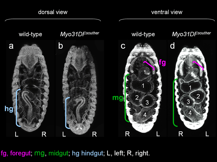
Mechanisms that create characteristic left-right asymmetry are largely unknown in invertebrate. To identify genes involved in left-right asymmetry of Drosophila embryonic gut, we performed a genetic screen. The embryonic gut is composed of three major parts, the foregut, midgut, and hindgut (a, c). We found that 75.7% of homozygous Myo31DFsouther embryos show synchronous inversion of the hindgut and midgut (b, d). In contrast, the handedness of the foregut was normal in all cases examined, indicating that this phenotype was heterotaxia (d). It is suggested that Myo31DF is crucial for generating left-right asymmetry of Drosophila embryonic gut. Myo31DF encodes an unconventional myosin, Drosophila ortholog of Myosin ID.
Shunya Hozumi, Reo Maeda, Kiichiro Taniguchi, Maiko Kanai, Syuichi Shirakabe, Takeshi sasamura, Pauline Spéder, Stéphane Noselli, Toshiro Aigaki, Ryutaro Murakami, Kenji Matsuno.
An unconventional myosin in Drosophila reverses the default handedness in visceral organs.
Nature 440, 798-802 (2006).
Runner-up
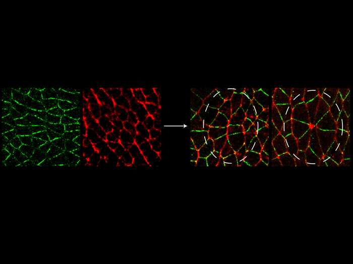
During germband extension in the Drosophila embryo, global planar polarities such as Bazooka (green) and Myosin II (red) produce locally coordinated cell rearrangements. Vertically arranged groups of cells contract internal cell interfaces to produce circular rosettes. These rosettes then resolve into horizontally elongated arrays, driving tissue elongation during convergent extension in the fly.
Blankenship JT, Backovic ST, Sanny JS, Weitz O, Zallen JA.
Multicellular rosette formation links planar cell polarity to tissue morphogenesis. Dev Cell 11:459-70 (2006).
Finalist
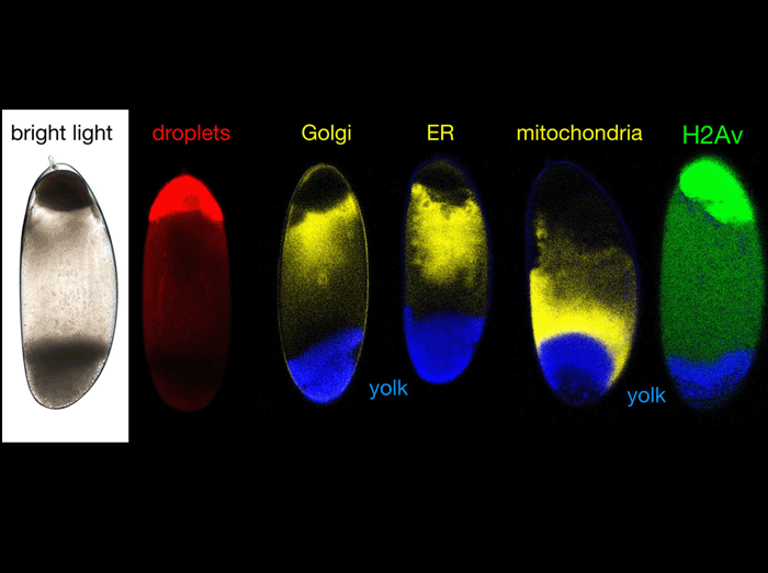
When living precellular embryos are oriented with their anterior end up and centrifuged, major organelles (lipid droplets, Golgi, ER, mitochondria and yolk) accumulate at distinct locations, resulting in consistent stratification in bright light. A GFP fusion of the histone variant H2Av is massively enriched in the droplet layer, revealing a surprising recruitment of certain histones to lipid droplets.
Silvia Cermelli, Yi Guo, Steven P. Gross, and Michael A. Welte
The lipid-droplet proteome reveals that droplets are a protein storage depot. Curr. Biol. 16:1783-1795 (2006)
Finalist
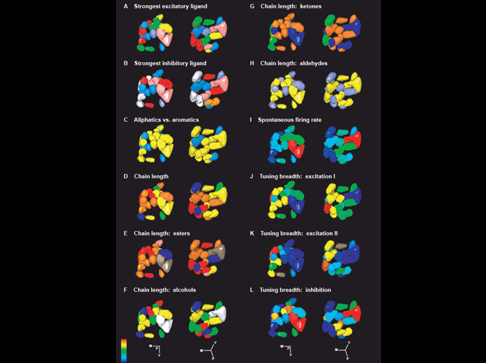
A central question in olfaction is how odorant responses are represented in the brain. In this figure, the response properties of odorant receptors to a panel of over 100 odors were measured and then mapped onto glomeruli in the antennal lobe. Each glomerulus is color-coded based on the response properties of the receptor expressed in its presynaptic ORN population. The figure shows that receptors with similar response properties often project to widely dispersed glomeruli, and receptors with very different response properties often project to neighboring glomeruli.
Elissa A. Hallem and John R. Carlson. 2006. Coding of odors by a receptor repertoire. Cell 125: 143-160.
Finalist
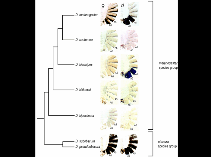
Body pigmentation is a rapidly evolving trait among Drosophila. Pigmentation of the posterior male abdomen recently evolved in the melanogaster species group. Frequent losses of male-specific pigmentation are observed in several lineages of themelanogaster species group, including D. santomea, D. kikkawai, and D. bipectinata. We infer that Abdominal-B Hox function has been co-opted to gain male-specific pigmentation at least in a common ancestor of the melanogaster species group. We further suggest three different mechanisms underlying the loss of male-specific pigmentation in different monomorphic lineages that descended from a common ancestor with male-specific pigmentation.
Sangyun Jeong, Antonis Rokas, and Sean B. Carroll.
Regulation of body pigmentation by the Abdominal-B Hox protein and its gain and loss in Drosophila evolution.
Cell 125, 1387-1399 (2006).
Finalist
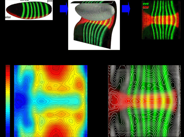
During the last mitotic interphase before gastrulation (stage 5), nuclei do not form three groups of distinct packing density as previously thought. Instead, the heat map on the bottom left shows a complex pattern of nuclear densities which varies continuously along both the a/p and d/v axes. The plot on the bottom right shows the same isodensity contours overlaid on the expression patterns of the genes snail (red) and even-skipped(green), revealing a complex relationship between gene expression and density. Both plots are cylindrical projections as explained on the top of the figure. Both the density and expression patterns have been averaged over 294 embryos.
Luengo Hendriks CL, Keränen SVE, Fowlkes CC, Simirenko L, Weber GH, DePace AH, Henriquez CN, Kaszuba DW, Hamann B, Eisen MB, Malik J, Sudar D, Biggin MD and Knowles DW.
3D morphology and gene expression in the Drosophila blastoderm at cellular resolution I: data acquisition pipeline
Genome Biology, 7:R123, 2006.
Finalist
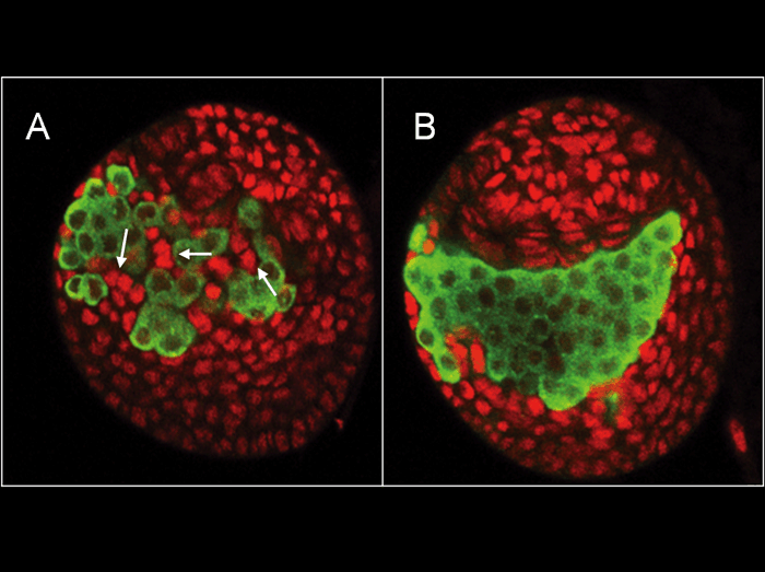
A. In Drosophila larval ovaries, somatic cells (red) and germ cells (green) interact and influence each other. A special group of somatic cells intermingles with germ cells (arrows). Intermingled cells and germ cells interact via a feedback loop containing a positive and a negative signal: The survival of Intermingled cells depends on Spitz, which is produced by germ cells. In return, Intermingled cells repress germ cell proliferation in an EGFR dependent manner.
B. Reducing EGFR signaling in somatic cells results in loss of Intermingled cells. This, in turn, increases the number of germ cells. The feedback loop is able to sense the number of germ cells and respond to keep their numbers within normal limits. It is therefore a homeostatic feedback loop that allows the organ to coordinate the growth of different cell populations.
Lilach Gilboa and Ruth Lehmann (2006)
Soma-germline interactions coordinate homeostasis and growth in the Drosophila gonad.
Nature, 443(7107):97-100.
Finalist
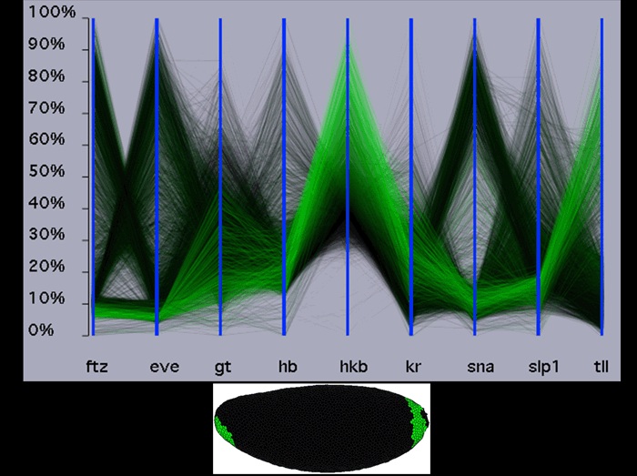
It is not possible to compare the expression of more than a handful of genes at once on a physical model of an embryo by using a different color to represent each gene because with too many genes the patterns overlap and colors mix. To overcome this limitation, we have adapted parallel coordinates, shown on the top panel, to represent and compare expression of multiple genes. In this parallel coordinates view, each gene is represented by a parallel vertical axis. The level of expression of one gene in one cell defines one point on a parallel axis. Connecting points for each cell on each axis results in zigzag lines, one zigzag line for each cell. In this example, the lines are colored according to the level of expression of the hkb gene. Below the parallel coordinate view, the locations of cells expressing hkb at the highest levels are shown on a physical model of the embryo. In the parallel coordinates, it can be readily seen that cells that express hkb at high levels express eve, ftz,sna and slp at low levels and tll at high levels. Although this illustration only compares nine gene’s expression, it is simple to display and compare data for tens of genes at once with further parallel vertical axes.
Rübel, O., Weber, G.H., Keranen, S.V.E., Fowlkes, C.C., Luengo Hendriks, C.L., Simirenko, L., Shah, N.Y., Eisen, M.B., Biggin, M.D., Hagen, H., Sudar, J.D., Malik, J., Knowles, D.W. and Hamann, B. (2006), PointCloudXplore: Visual analysis of 3D gene expression data using physical views and parallel coordinates, in: Sousa Santos, B., Ertl, T. and Joy, K.I., eds., Data Visualization 2006 (Proceedings of EuroVis 2006), Eurographics Association, Aire-la-Ville, Switzerland, pp. 203-210.
