2019 Drosophila Image Award
Winner – Video
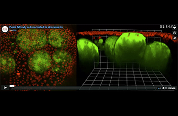
Pupal fat body cells recruited to skin wounds
Pupal fat body cells are motile cells that are recruited to skin wounds. This movie shows one fat body cell approaching an epithelial wound and forming a tight bond with the wound gap until wound re-epithelialisation is complete. Other fat body cells appear later and are seen patrolling in the vicinity. Fat body cells are shown in green (c564-Gal4+UAS-GFP) and epithelial nuclei in red (Ubq>Histone-RFP). Projection and Z plane views are shown on the left and right, respectively. Elapsed time is in top right corner in hours:minutes:seconds. Scale bar, 20 μm
Anna Franz, Will Wood and Paul Martin (2018)
Fat body cells are motile and actively migrate to wounds to drive repair and prevent infection.
Dev Cell. 2018 Feb 26; 44(4):460-470.e3
Winner – Still Image
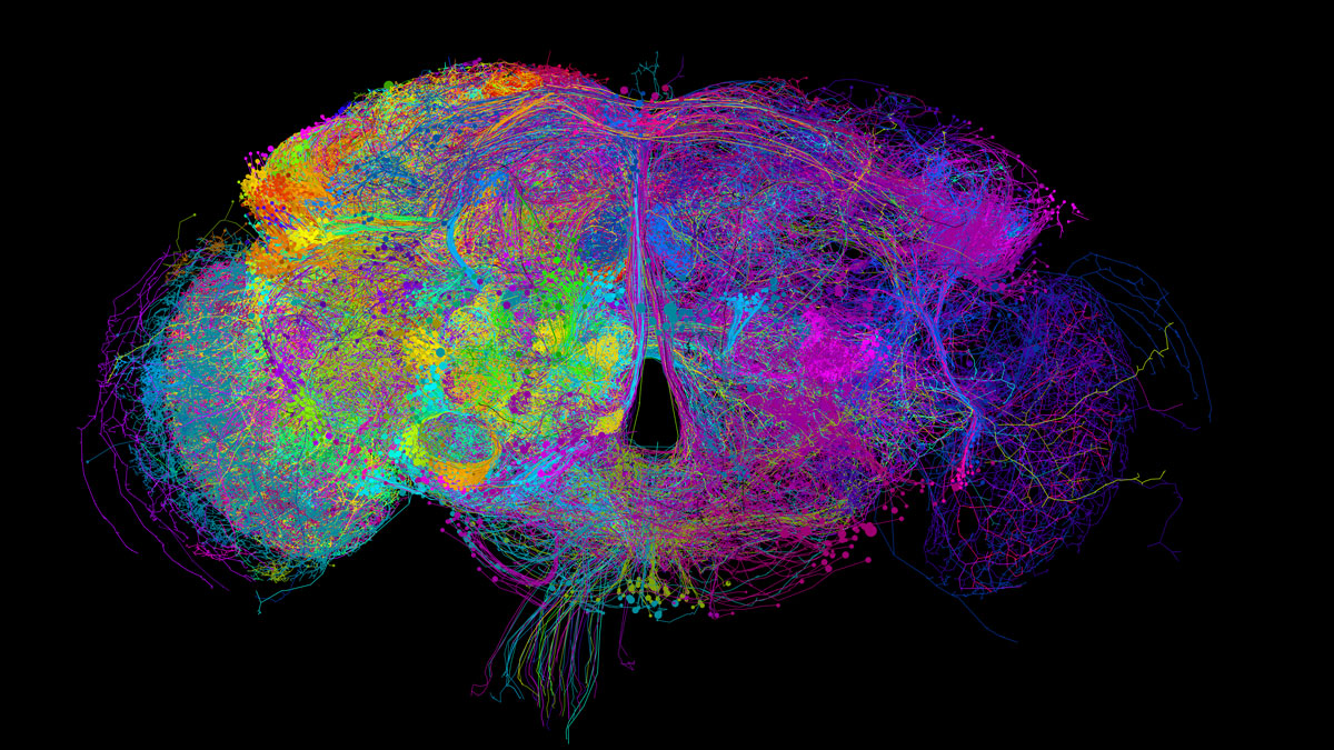
Reconstruction of Neurons from an Electron Microscopy Volume of the Entire Adult Fruit Fly Brain
The neural basis for behavior and decision-making is defined, in part, by the wiring diagram between neurons in the brain. Zheng, Lauritzen et al. present a whole-brain electron microscopy image dataset of an adult fruit (or vinegar) fly, Drosophila melanogaster, sufficient to trace the synaptic connectivity of any selected brain circuit in this genetic model organism. In the image, a reconstruction of neurons from the volume are colored according to their soma locations. Over 20 labs have collectively reconstructed ~7 meters of cable length, ~5% of total in brain.
EM reconstruction of Drosophila neurons by the Full Adult Fly tracing community.
Image Credit: Philipp Schlegel (Drosophila Connectomics Group, Cambridge)
Zheng Z, Lauritzen JS, Perlman E, Robinson CG, Nichols M, Milkie D, Torrens O, Price J, Fisher CB, Sharifi N, Calle-Schuler SA, Kmecova L, Ali IJ, Karsh B, Trautman ET, Bogovic JA, Hanslovsky P, Jefferis GSXE, Kazhdan M, Khairy K, Saalfeld S, Fetter RD, Bock DD (2018)
A Complete Electron Microscopy Volume of the Brain of Adult Drosophila melanogaster.
Cell. 2018 Jul 26;174(3):730-743
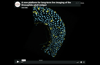
A new platform for long-term live imaging of the Drosophila adult midgut
The midgut of adult Drosophila has emerged as a premier genetic model for stem cell biology. However, its potential has been constrained by the lack of a robust imaging methodology. A new platform for imaging live Drosophila adults (top panel) over prolonged time scales opens the door to real-time study of organ renewal in a near-native context. A window cut into the dorsal cuticle (bottom panel) allows the midgut to be imaged while the animal continues to eat, undergo peristalsis, and defecate. A 14.8-hour time series of a peristalsing midgut tube (right video) illustrates the dynamic, high-resolution image data that are captured with this approach. By enabling real-time study of midgut phenomena that were previously inaccessible, our platform opens a new realm for dynamic understanding of adult organ renewal.
Cyan, all nuclei (ubi-his2ab::mRFP) ; yellow, midgut stem cell and enteroblast progenitors (esgGAL4; UAS-LifeactGFP).
J. Martin, E. Sanders, P. Moreno-Roman, L. Koyama, S. Balachandra, X. Du, & L.E. O’Brien. (2018)
Long-term live imaging of the Drosophila adult midgut reveals real-time dynamics of division, differentiation and loss.
eLife 7:e36248 DOI: 10.7554/eLife.36248
1st Runner Up
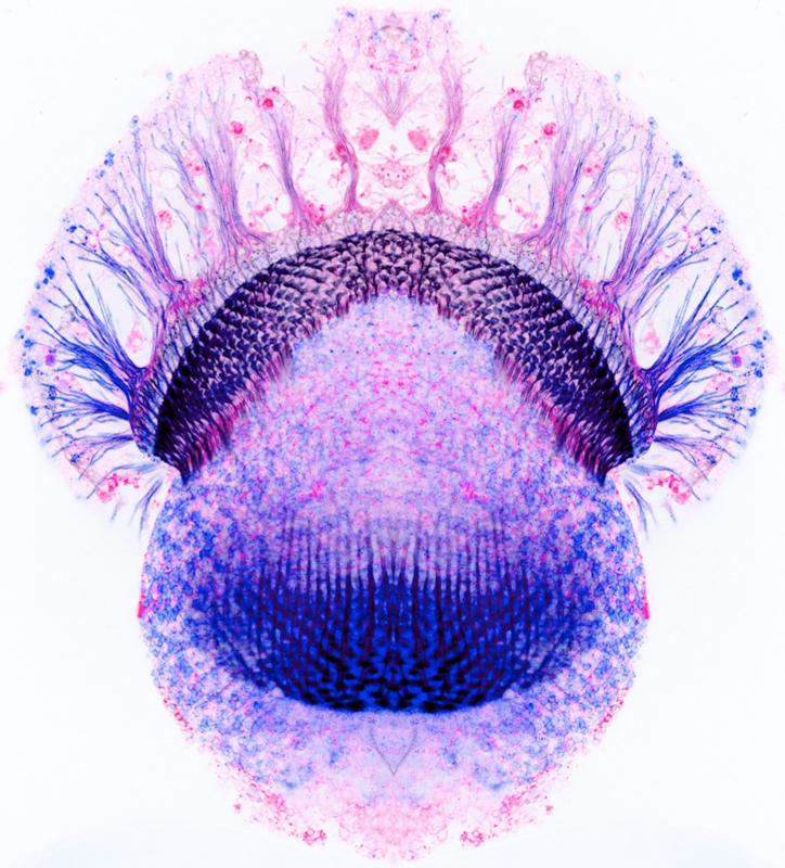
Splicing reporters reveal cell-type-specific isoform expression
The confocal image shows the expression of Dscam2 isoform B expression in the Drosophila optic lobe during mid-pupal development. The left and right sides are mirrored to provide aesthetic symmetry and the colors are inverted to highlight the complexity of neuronal projections within the brain.
The alternative splicing of the cell adhesion molecule, Dscam2, is neuronal cell-type-specific. This permits the proper development of axon terminals of tightly associated neurons. Our study further showed that this deterministic splicing is necessary for attaining the appropriate number of synapses and the morphology of dendrites. These results provide insight into how functional diversity is achieved by the genome.
Sarah K. Kerwin*, Joshua S.S. Li*, Peter G. Noakes, Grace J. Shin and S. Sean Millard (2018) (*equal contribution)
Regulated Alternative Splicing of Drosophila Dscam2 Is Necessary for Attaining the Appropriate Number of Photo
Honorable Mention
Optically-cleared GFP-positive D. melanogaster from larva to adult recorded with light-sheet microscopy
Imaging of cells or molecules in the context of their natural tissue environment in undissected D. melanogaster has been limited due to different pigments and optical properties of the cuticle and/or the pupal case.
So far sample preparation is required including the generation of histological sections or the dissection of organs before mounting and imaging. In either case this leads to tissue damage, deformation and loss of information especially in cases like the nervous system, where dissected nerves and individual neuronal projections are notoriously difficult to reconstruct.
We developed a clearing technique for whole D. melanogaster from larva to adult. During this process, pigments from the body and the compound eye are removed while the signal of several endogenously expressed fluorophores is preserved. Combined with light-sheet microscopy, this enables visualization of e.g. whole nervous system.
Shown are whole mount recordings of Peb-Gal4 UAS-CD8::GFP larva, prepupa, pupa and adult fly (from top to down) and olfactory and visual system in adult D. melanogaster head.
Video Credit: Marko Pende
Pende, M., Becker, K., Wanis, M., Saghafi, S., Kaur, R., Hahn, C., Pende, N., Foroughipour, M., Hummel, T., and Dodt, H.-U.
High-resolution ultramicroscopy of the developing and adult nervous system in optically cleared Drosophila melanogaster.
Nat Commun 9, 4731. (2018)
Honorable Mention
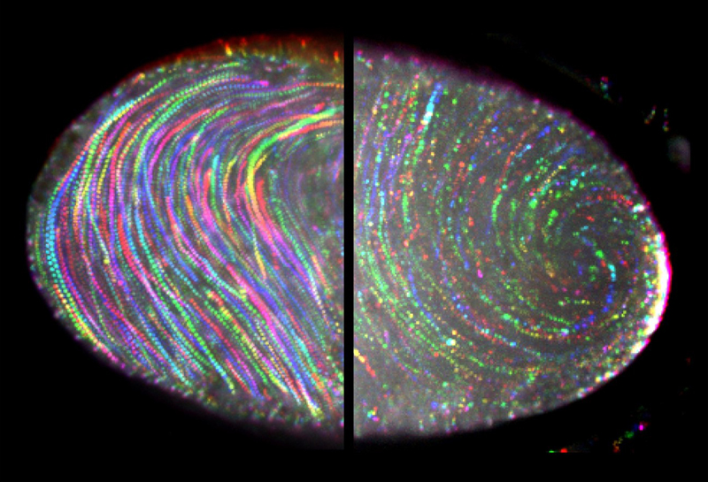
Ooplasmic streaming
During late oogenesis, Drosophila oocyte cytoplasm undergoes circular movement called ooplasmic streaming. Posterior determinants containing the fluorescently labeled marker Staufen move with the cytoplasmic flow, but dramatically slow down near the posterior pole in a wild-type oocyte (right) but not in an oocyte expressing a dominant-negative form of myosin V (left). Spectrum-colored tracks represent the movement of Staufen-containing particles within a 10-min period of time.
Wen Lu, Margot Lakonishok, Anna S. Serpinskaya, David Kirchenbüechler, Shuo-Chien Ling, Vladimir I. Gelfand (2018)
Ooplasmic flow cooperates with transport and anchorage in Drosophila oocyte posterior determination.
Journal of Cell Biology. 217(10):3497-3511
Honorable Mention
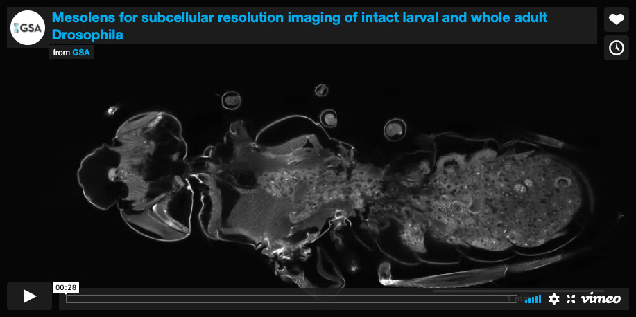
Mesolens for subcellular resolution imaging of intact larval and whole adult Drosophila
Confocal Mesolens z-series and rotating 3D view of whole female Drosophila imago with sub-cellular resolution throughout the first part of the movie shows a z-series of 242 images through a whole female Drosophila imago, with all external and internal structure visible. The second part of the movie shows the exterior of the fly in a rotating 3D reconstruction of the same specimen, created using the ‘3D Rotation’ plugin in the image processing software Icy. A series of projections at different angles (2 degrees, through 360 degrees) was made using a maximum brightness projection algorithm, which effectively eliminated all but the bright exoskeletal signal. We note that unlike projections of z-series made with conventional microscope methods, projections of our Mesolens images do not show blurring in the optically axial direction (here used as the rotation axis). This is because the axial resolution length is small compared with the height of the specimen.
G. McConnell and W.B. Amos. (2018)
Application of the Mesolens for subcellular resolution imaging of intact larval and whole adult Drosophila
J. Microscopy, Vol. 270, Issue 2 2018, pp. 252–258
Honorable Mention
Social learning of mating preferences in Drosophila
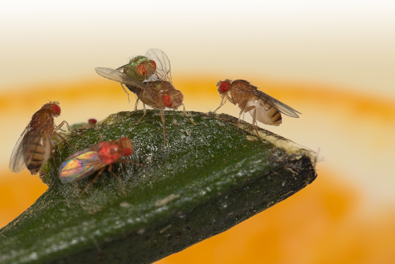
A situation of mate-copying in which two females watch a copulating green male while a pink male is rejected.
Image Credit: Duneau David
Etienne Danchin, Sabine Nöbel, Arnaud Pocheville, Anne-Cecile Dagaeff, Léa Demay, Mathilde Alphand, Sarah Ranty-Roby, Lara van Renssen, Magdalena Monier, Eva Gazagne, Mélanie Allain, Guillaume Isabel (2018)
Cultural flies: Conformist social learning in fruitflies predicts long-lasting mate-choice traditions
Science 362, 1025–1030
Honorable Mention
One lucky germ cell
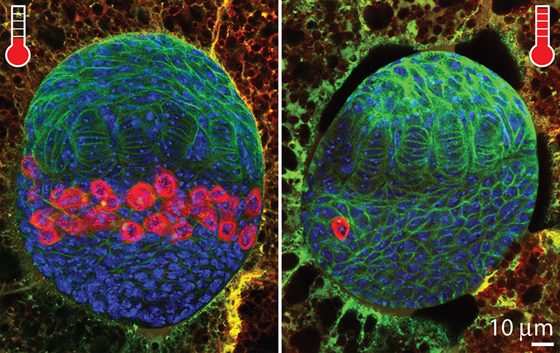
Given the ability to jump around the genome, transposon invasion often leads to animal sterility. By simply adjusting temperature that modulates the activity of P-elements (a DNA transposon), we were able to observe the initial response from germline stem cells that lead to a robust transposon-endogenizing process. Shown is the consequence of transposon invasion in germ cells (red) at 3rd instar larvae stage. Compared to modest transposon activity (left panel; low temperature), strong transposon activity (right panel; high temperature) results in severe loss of the germ cells that subsequently develops rudimentary ovary in adult stage.
Sungjin Moon, Madeline Cassani, Yu An Lin, Lu Wang, Kun Dou, ZZ Zhao Zhang (2018)
A robust transposon-endogenizing response from germline stem cells.
Developmental Cell 47(5), 660-671 (2018).
Honorable Mention
Drosophila imaged with the Mesolens
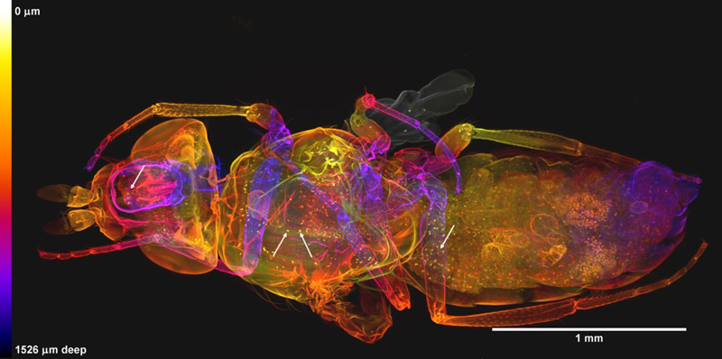
Whole female Drosophila newly emerged imago in dorsal view imaged with the Mesolens. This image is composed by projection of 242 optical sections taken with an axial separation of 6.3 μm, forming a z-stack 1.53 mm deep. The sections are colour-coded for depth according to the scale shown, in which near sections are yellow and the far ones purple or dark grey. Only propidium iodide (PI) was used as a fluorochrome, revealing both cuticle and nuclei of individual cells in the interior. The orange clusters of nuclei in the abdomen are those of the ovaries (indicated with large white arrows at the right-hand side of the image). The smaller white arrows on the left of the image indicate PI-positive bodies that may be parasites. This dataset is representative of n=7 adult flies imaged using the same method.
G. McConnell and W.B. Amos. (2018)
Application of the Mesolens for subcellular resolution imaging of intact larval and whole adult Drosophila
J. Microscopy, Vol. 270, Issue 2 2018, pp. 252–258
Honorable Mention
Drosophila species learn dialects through communal living
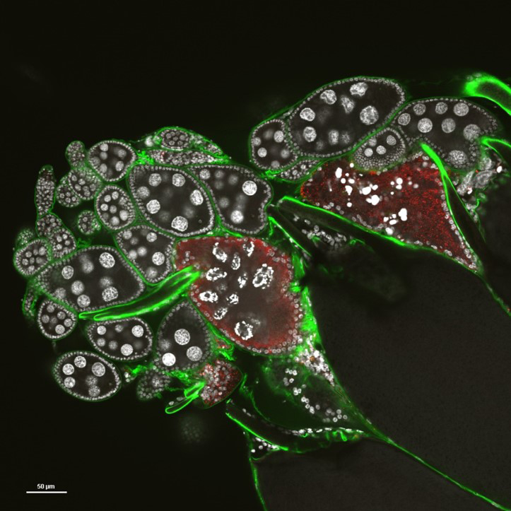
A light microscope image of dissected Drosophila ovary where DNA (white) reveals perfectly round nurse and follicle cell nuclei of intact eggchambers in addition to bright-staining, condensed fragmented DNA from apoptotic nurse cells expressing activated caspases (red). The outline of egg surfaces is labeled by wheatgerm agglutinin (green). Predatory wasps inject their eggs into Drosophila larvae, but adult female flies can see the predator, or simply be informed by other flies exposed to wasp through social learning. Subsequent to a visual cue from the wasp or an informed teacher, flies deprive the predator of larvae by triggering death of its own oocytes and halting egg production.
Kacsoh BZ, Bozler J, Bosco G (2018)
Drosophila species learn dialects through communal living.
Plos Genetics
Honorable Mention
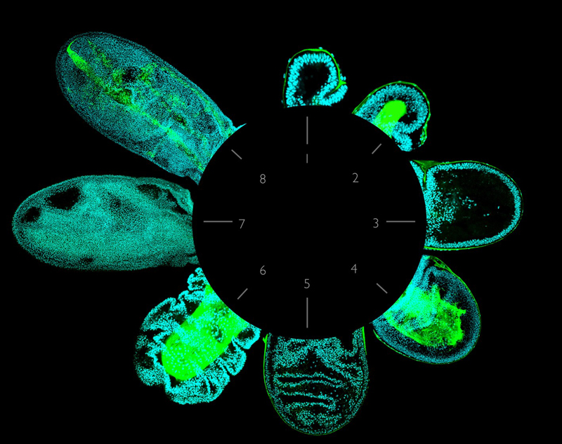
From Wings to Halteres
In dipteran insects such as Drosophila, the anterior pair of wings are used for flight, while the posterior pair are dramatically reduced to form a balancing structure called the haltere. The key factor that controls this difference is the Homeobox gene Ultrabithorax. We have shown that one of the main ways in which Ultrabithorax performs its task is by inhibiting the degradation of both apical (aECM) and basal extracellular (bECM) matrixes -a process that normally occurs in the wing at the beginning of metamorphosis- thus impairing elongation and the flattening of the haltere.
The picture shows how the shape of Drosophila appendages at early metamorphosis (nuclei, cyan) depend on ECM localisation (green): Halteres (1, aECM; 2, bECM) and wings (3, aECM; 4, bECM), before ECM degradation in the wing is due; mutant wings in which either the apical (5) or the basal (6) ECM cannot be degraded; and elongated and expanded wings after ECM degradation (7, aECM; 8, bECM).
Maria-del-Carmen Diaz-de-la-Loza, Robert P. Ray, Poulami Somanya-Ganguly, Silvanus Alt, John R. Davis, Andreas Hoppe, Nic Tapon, Guillaume Salbreux and Barry J. Thompson. (2018)
Apical and Basal Matrix Remodeling Control Epithelial Morphogenesis.
Cell, July 2 2018. 46, 23-39 2018 46, 23-39.
Honorable Mention
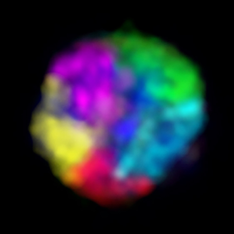
Chromosome territory formation in a Drosophila interphase nucleus
During interphase, chromosomes in all eukaryotic species are partitioned into distinct domains called chromosome territories (CTs). Here, we took advantage of the relatively small genome of Drosophila melanogaster to visualize CT formation genome-wide using the Oligopaints technology.
This figure not only demonstrates how Oligopaints can be scaled to make efficient multi-color chromosome paints, but also illustrates that Drosophila embryonic nuclei have robust CT formation. Shown here: Oligopaints labeling all the unique sequences of chromosome X in green, 2L in red, 2R in cyan, 3L in yellow, and 3R in magenta.
Leah F. Rosin, Son C. Nguyen, Eric F. Joyce. (2018)
Condensin II drives large-scale folding and spatial partitioning of interphase chromosomes in Drosophila nuclei.
PLOS Genetics 14(7): e1007393
Honorable Mention
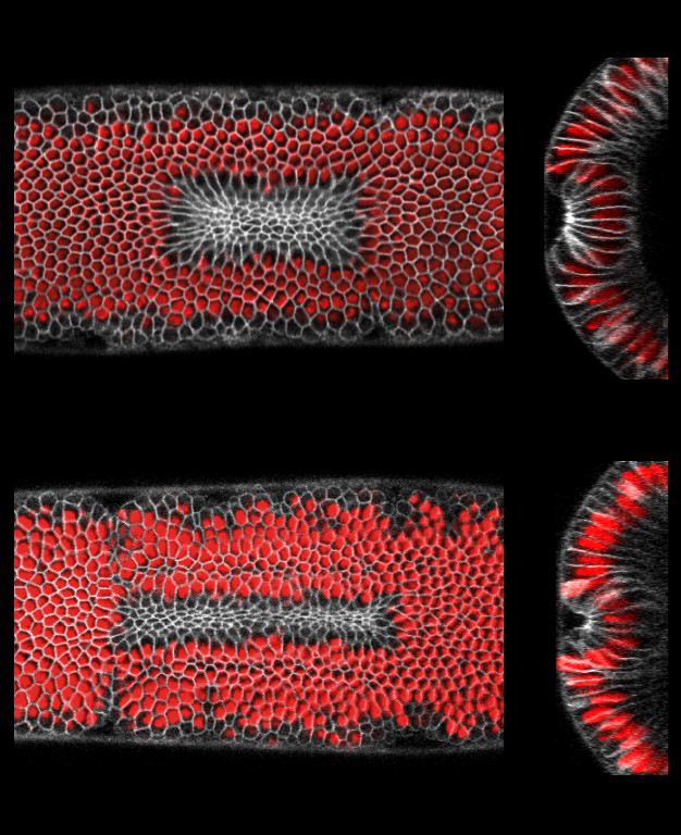
Synthetic furrows through optogenetic activation of cell contractility
Morphogenesis starts with local changes in cell shape that are translated into changes a the tissue level. In turn, changes in the tissue also feedback to the cell level. This interplay is key for the coordination and robust execution of development, however, how this interplay occurs is poorly understood. We used optogenetics to control the spatial and temporal activation of Rho signaling. We show that activation of Rho1 is sufficient to trigger apical constriction and tissue folding. Moreover, we show that individual cells within activated areas of different rectangularity (top and bottom) respond differently to tissue level cues (e.g. tissue geometry). Cells elongate as the geometry of the contractile domain increases in rectangularity, and the elongation of cells orients parallel to the longer axis of the activated region. These observations demonstrate that geometrical constraints provide a tissue level cue for cell and tissue shape changes.
Emiliano Izquierdo, Theresa Quinkler, Stefano De Renzis. (2018)
Guided morphogenesis through optogenetic activation of Rho signalling during early Drosophila embryogenesis.
Nature Communications. Jun 18; 9(1):2366
