2023 Drosophila Image Award
Winner – Video
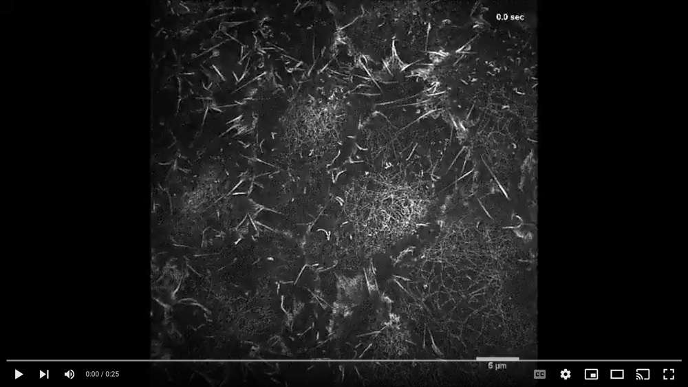
Grazing Incidence, Structured Illumination Microscopy demonstrates that networks of individual, fluorescent actin filaments condense and relax in cells undergoing pulsed contractions and expansions during dorsal closure
Pulsed cellular contractions and expansions occur on a 10s of second time scale occur midway through embryogenesis in Drosophila melanogaster – ultimately, they contribute to emergent properties that contribute to tissue morphogenesis on a tens of minutes (hours) time scale. The embryos were fluorescently labeled through the ubiquitous expression of GFP-fused to the actin binding domain of fly moesin. The time-lapsed sequence is sped up 30 times. The field of view of the movie is larger than the region of interest shown in Figure 1 of the publication that fully describes this work:
Regan P. Moore, Stephanie M. Fogerson, U. Serdar Tulu, Jason W. Yu, Amanda H. Cox, Melissa A. Sican, Dong Li, Wesley R. Legant, Aubrey V. Weigel, Janice M. Crawford, Eric Betzig, and Daniel P. Kiehart.– Stills from the movie are shown in Figure 1 and at high magnification in Figures 2 and 4 of the paper.
Video Credit: Regan Moore, U. Serdu Tulu, Dong Li
Moore RP, Fogerson SM, Tulu US, Yu JW, Cox AH, Sican MA, Li D, Legant WR, Weigel AV, Crawford JM, Betzig E, Kiehart DP.
Superresolution microscopy reveals actomyosin dynamics in medioapical arrays.
Mol Biol Cell. 2022 Sep 15;33(11):ar94.
Winner – Still Image
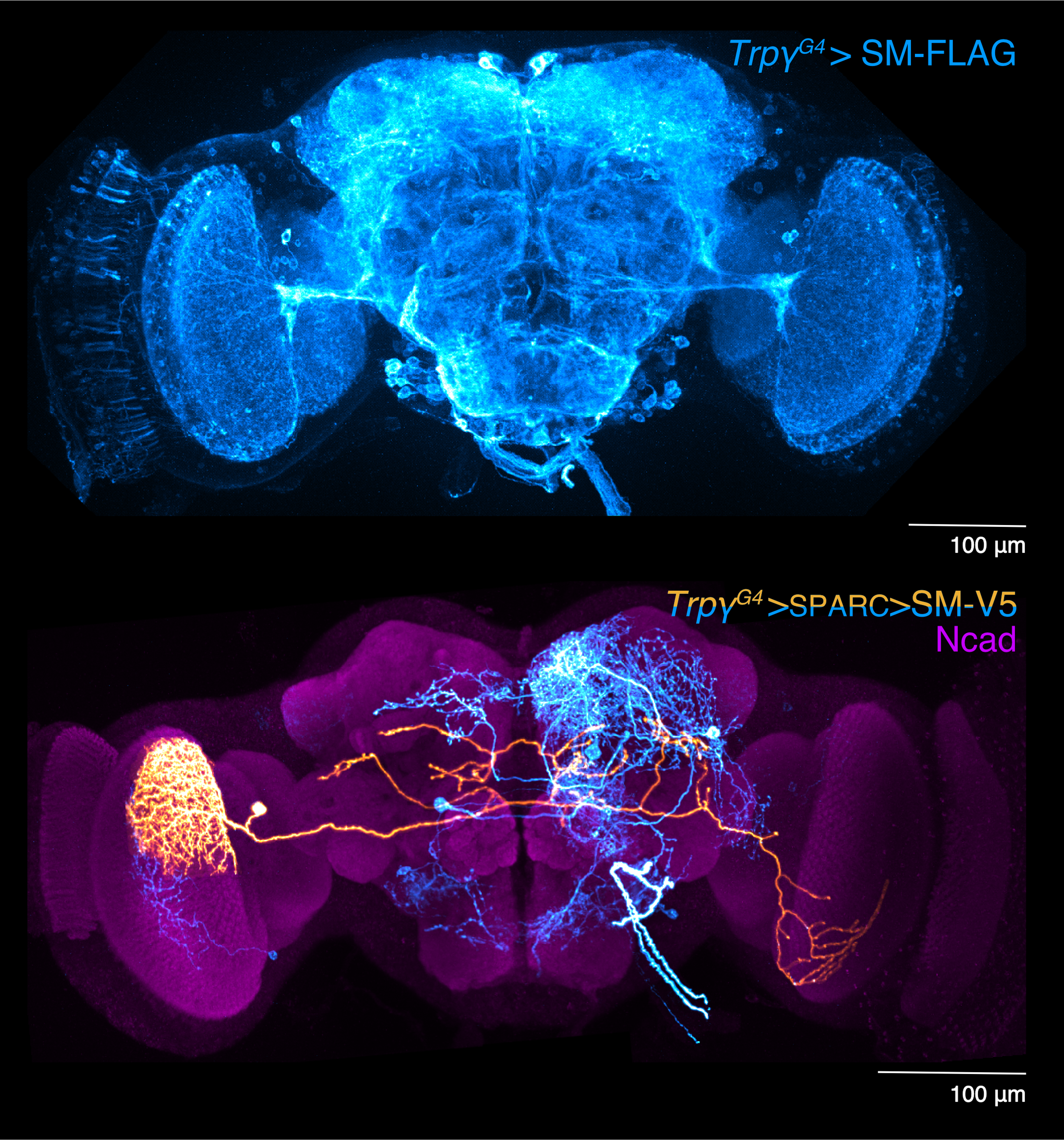
Arborizations of Trpγ+ neurons, a developmental neuronal population that coordinates brain-wide activity
Patterned, stimulus-independent neural activity (PSINA) propagates throughout the brain during Drosophila pupal development. Approximately 2000 neurons express the cation channel transient receptor potential gamma (Trpγ). These neurons arborize throughout the brain where they relay and pattern PSINA. The panel above shows coverage of the brain 72 hours after pupal formation by Trpγ+ neurons expressing myr::SM-FLAG. Most Trpγ+ neurons are labeled; some sparseness was introduced using FLP-Out to improve staining. The panel below highlights a single Trpγ+ neuron (orange, manually segmented) in the context of others (cyan) labeled using Sparse Predictive Activity through Recombinase Competition (SPARC). Both images are maximum intensity projections of a confocal stack.
Image Credit: Bryce Bajar and Orkun Akin
Bajar BT, Phi NT, Isaacman-Beck J, Reichl J, Randhawa H, Akin O.
A discrete neuronal population coordinates brain-wide developmental activity.
Nature. 2022 Feb; 602(7898), 639-646.
1st Runner Up – Video
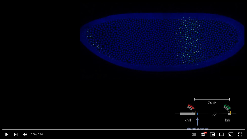
Real-time visualization of endogenous transcription of knirps (kni) and knirp-related (knrl), during nuclear cycle 14
Despite 74kb separation the genes display concurrent activities in space and time, as well as co-initiation events (<90 seconds) in single nuclei. A shared enhancer, proximal to knrl and distant from kni, underlies co-regulation in the anterior stripe domain. Regulatory interconnectivity further depends on promoter-proximal tethering elements, and perturbations in these elements uncouple transcription and alter the bursting dynamics of the distant gene. Transcriptional coupling is detected throughout the fly genome and encompasses a broad spectrum of conserved developmental processes, suggesting a general strategy for long-range integration of gene activity. Video Credit: Michal Levo, João Raimundo Levo M , Raimundo J, Bing XY, Sisco Z, Batut PJ, Ryabichko S, Gregor T, Levine MS.
Transcriptional coupling of distant regulatory genes in living embryos.
Nature. 2022 May; 605(7911), 754-760.
1st Runner Up – Still Image
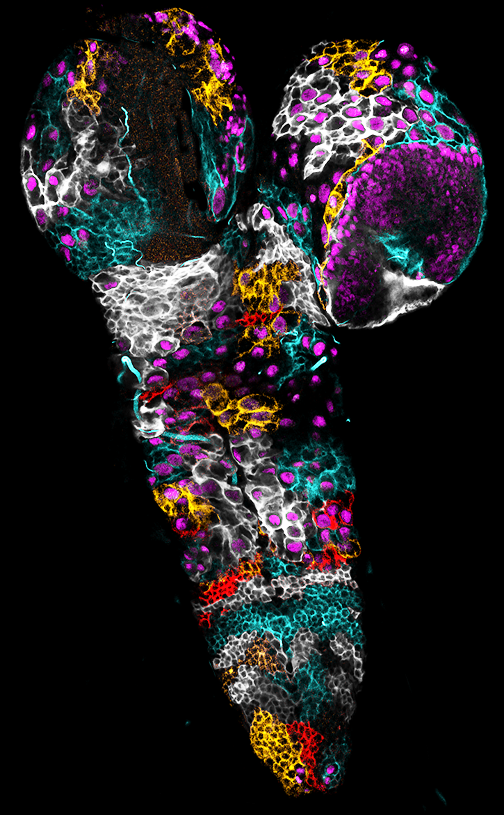
Mosaic of cortex glia cells in the larval central nervous system, revealed by multicolour membrane labelling
Cortex glia form a membrane network spanning the whole larval central nervous system, while encasing individual neural stem cells in a bespoke chambre. Stochastic multicolour labelling (membrane-tagged Raeppli-CAAX: White, Cyan, Orange and Red) revealed the contribution of individual cells to this structure. First, individual cortex glia cells encase several neural stem cells (nuclei in magenta), partitioning the stem cell population in spatial modules. Moreover, while they tile the organ in mostly non-overlapping domains, cortex glia are able of homotypic cell-cell fusion, remodelling the boundaries of these modules.
Image Credit: Pauline Spéder
Rujano MA, Briand D, Ðelić B, Marc J, Spéder P.
An interplay between cellular growth and atypical fusion defines morphogenesis of a modular glial niche in Drosophila.
Nat Commun. 2022 Aug 25; 13(1), 4999.
2nd Runner Up – Video
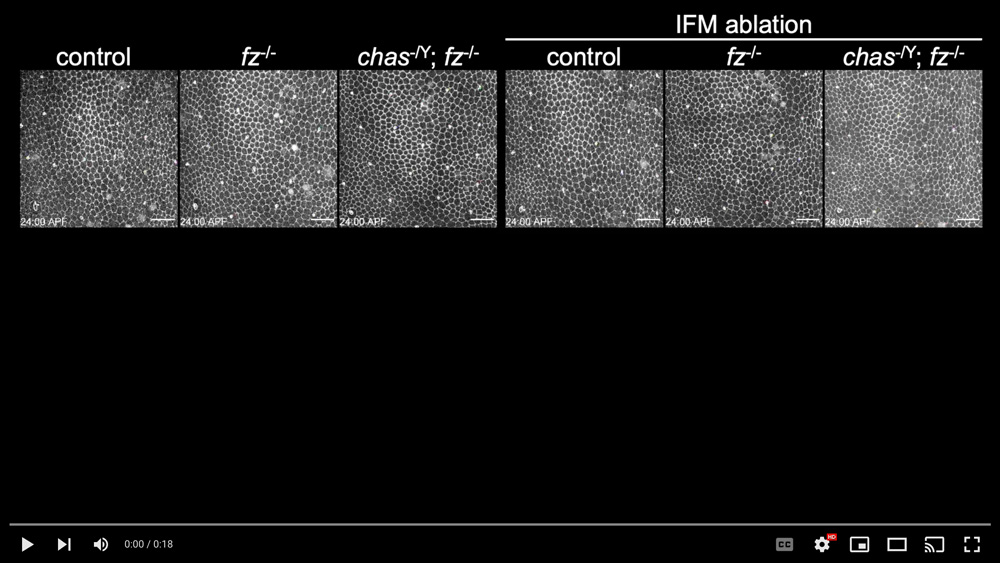
Tissue flow can orient bristles in the opposite direction to the flow
Planar cell polarity (PCP) regulates the orientation of external structures. A core group of proteins that includes the transmembrane protein Frizzled forms the heart of the PCP regulatory system. Other PCP mechanisms that are independent of the core group likely exist but their underlying mechanisms are unclear. We found that tissue flow is a mechanism governing core group-independent PCP on the Drosophila notum and can orient bristles in the opposite direction to the flow. This movie shows that tissue flow manipulation can change notal bristle orientation. A central notum region anterior to the aDC bristle (the white box in leftmost panel in the middle low) was analyzed. The anterior is at the top and the posterior is at the bottom. Top panels: Live imaging of tissue flows in control, fz mutant and chas; fz double mutant nota expressing E-cadherin::GFP without and with indirect flight muscle (IFM) ablation. Time (h:min). Scale bar, 20 μm. Middle panels: Macroscopic dorsal views of notal bristle phenotypes in adult control flies and flies deficient in the indicated PCP genes, as well as control flies and the same mutant strains subjected to IFM ablation. Bottom panels: Rose diagrams showing the angular distribution of the bristles.
Video Credit: Tomonori Ayukawa, Masakazu Yamazaki
Ayukawa T, Akiyama M, Hozumi Y, Ishimoto K, Sasaki J, Senoo H, Sasaki T, Yamazaki M.
Tissue flow regulates planar cell polarity independently of the Frizzled core pathway.
Cell Rep. 2022 Sep 20; 40(12):,111388.
2nd Runner Up – Still Image
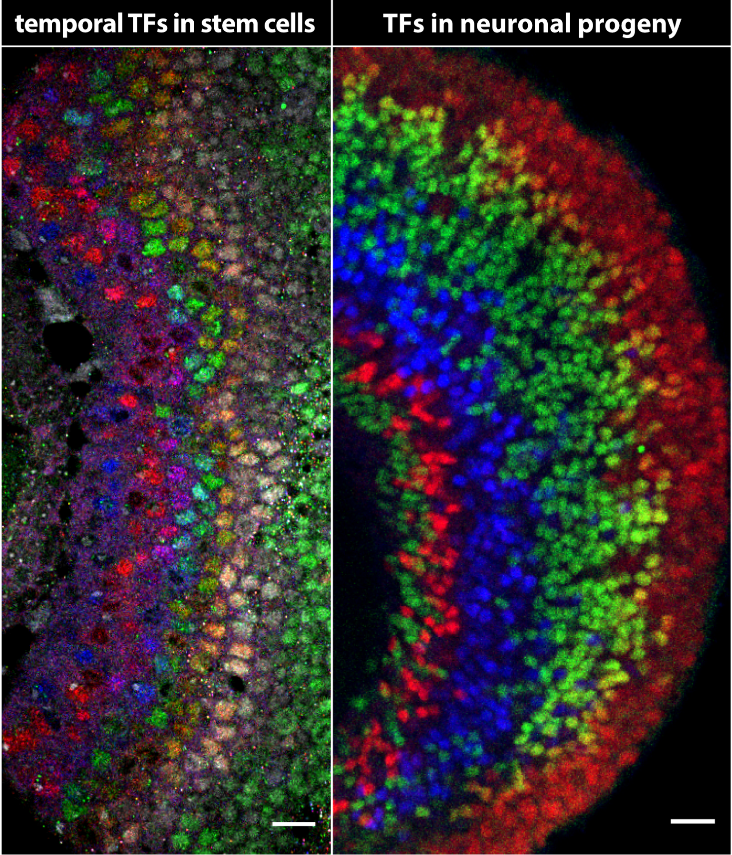
Temporal patterning of Drosophila neural stem cells generates neural diversity over time
Temporal patterning is a neuronal specification mechanism by which neural stem cells sequentially express different transcription factors (termed temporal TFs) as they age to generate different neuronal types over time. The left panel shows previously uncharacterized temporal TFs sequentially expressed in medulla stem cells, while the right panel shows early to late born medulla neurons that express different neuronal TFs and are generated from these neuroblasts.
Image Credit: Isabel Holguera
Konstantinides N, Holguera I, Rossi AM, Escobar A, Dudragne L, Chen YC, Tran TN, Martínez Jaimes AM, Özel MN, Simon F, Shao Z, Tsankova NM, Fullard JF, Walldorf U, Roussos P and Desplan C.
A complete temporal transcription factor series in the fly visual system.
Nature. 2022 Apr; 604(7905), 316-322.
Honorable Mention – Video
Injured Dendrites Exhibit Calcium-Flashing
For long-term lapse live imaging, 3rd instar Drosophila larvae expressing myr-GCaMP6s (green) and CD4-tdTom (magenta) in class IV dendritic arborization neurons were mounted using LarvaSPA technique. Dendrites were precisely ablated at a branching point near the cell body by two-photon laser. Continuous time-lapse imaging revealed that severed dendrites exhibit fast calcium flashing, which lasts for hours, before the dendrites enter a quiescence phase and finally fragment with a calcium surge. This calcium flashing is absent in severed axons and may be important for triggering dendrite-specific degeneration programs.
Video Credit: Hui Ji
Ji H, Sapar ML, Sarkar A, Wang B, Han C.
Phagocytosis and self-destruction break down dendrites of Drosophila sensory neurons at distinct steps of Wallerian degeneration.
PNAS 2022 Jan 20; 119(4), e2111818119.
Honorable Mention – Still Image
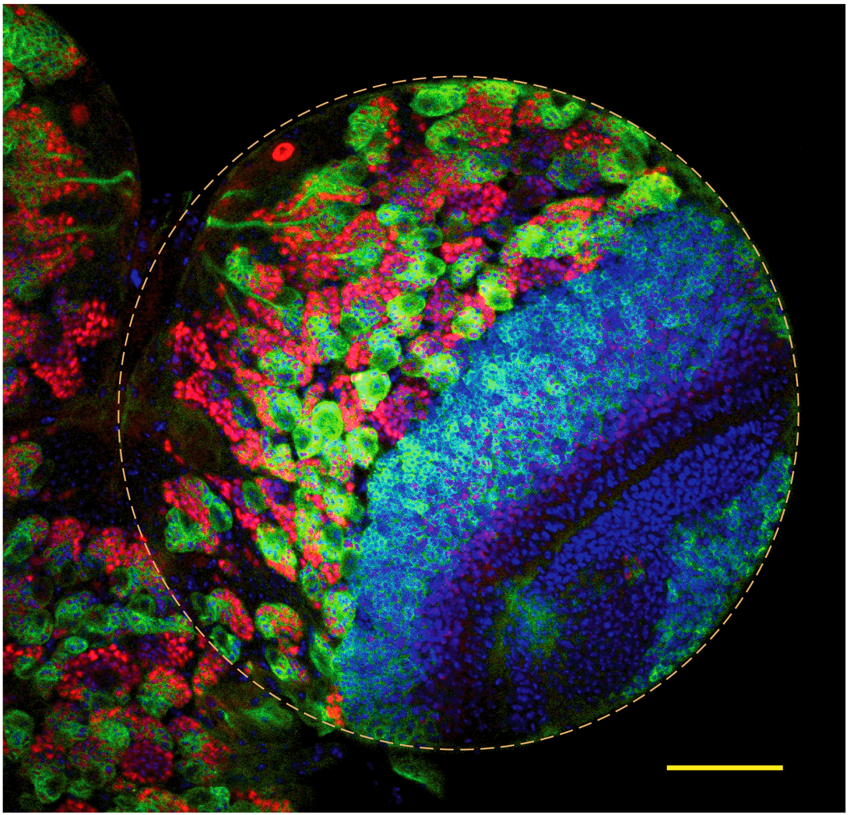
Neural stem cell lineages in the Drosophila larval brain
The fluorescence confocal image shows Drosophila larval brain hemisphere (yellow dotted circle) containing numerous neural stem cell (NSC) lineages marked with mCD8-GFP (green). Each lineage can be identified by the presence of large and round NSC at the top surrounded by its daughter neurons (red) marked by nuclear Prospero. All nuclei in the brain are marked by the hoescht staining. Scale bar- 50um
Image Credit: Dnyanesh Dubal
Dubal D, Moghe P, Verma RK, Uttekar B, Rikhy R.
Mitochondrial fusion regulates proliferation and differentiation in the type II neuroblast lineage in Drosophila.
PLoS Genet 2022 Feb 14; 18(2), e1010055.
Honorable Mention – Video
Imaging of Larval Eye Imaginal Disc Development Ex Vivo.
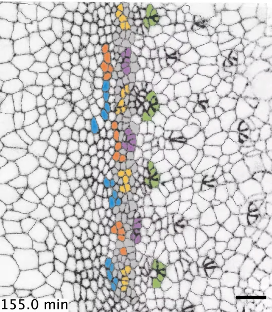
This video of ten hours development shows five columns of ommatidia emerge from the morphogenetic furrow as it moves across the eye disc. All presumptive R2, R3, R4, R5, and R8 cells belonging to the same column are labelled with the same color. Cells shaded grey represent the stripe of cells residing within the furrow. Note that cells in the furrow including presumptive R cells flow with the furrow until they stop flowing and are left behind.
Video Credit: Kevin Gallagher and Richard W. Carthew
Gallagher KD, Mani M, Carthew RW.
Emergence of a geometric pattern of cell fates from tissue-scale mechanics in the Drosophila eye.
eLife 2022 Jan 17; 11, e72806.
Honorable Mention – Still Image
Inter-tissue cooperation for tissue gap closure
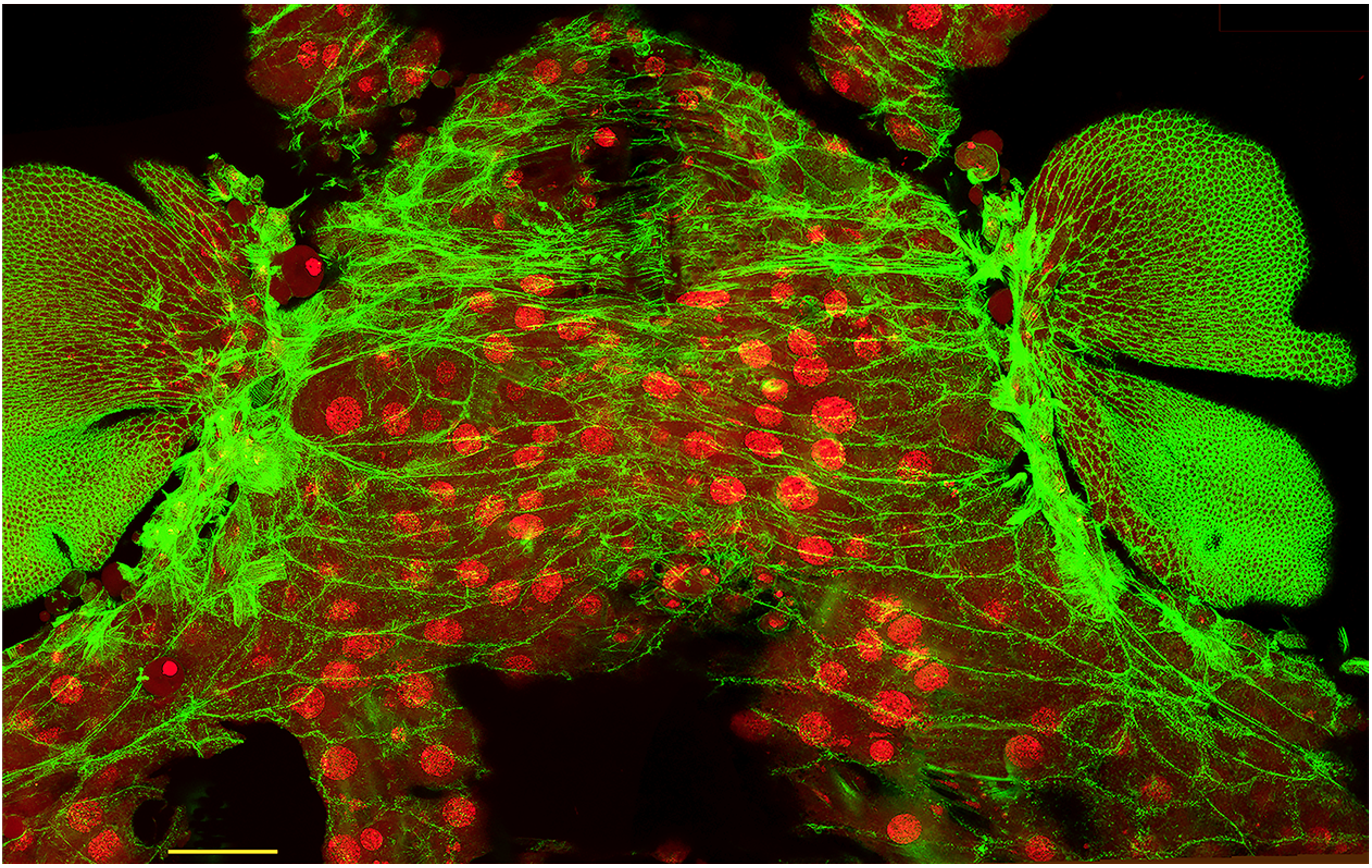
This image represents a snapshot of Drosophila pupal thorax closure, where two contralateral heminotal epithelia (HE) migrate over an underlying larval epidermal cell (LEC) layer. Both these cell layers are visualized by actin (green), while the latter further displays large polyploid nuclei (red). How these two HEs migrate over a distance nearly thrice their respective sizes has been a riddle. We show that the LEC layer generates contraction forces—as suggested by the formation of actin stress fibers (green)—pulling the contralateral HEs together for thorax closure thereby forming a bilaterally symmetrical adult thorax. Our findings reveal the crucial role of a degenerating cell layer in tissue gap closure during organogenesis. This mechanism may recur during wound healing as well. Scale bar: 50 µm.
Image Credit: Saurabh Singh Parihar and Rachita Bhattacharya.
Athilingam T, Parihar SS, Bhattacharya R, Rizvi MS, Kumar A, Sinha P.
Proximate larval epidermal cell layer generates forces for pupal thorax closure in Drosophila.
Genetics 2022 May 5; 221(1), iyac030.
Honorable Mention – Video
Multiscale 360° view of a living Drosophila pupa imaged by MuViScopy
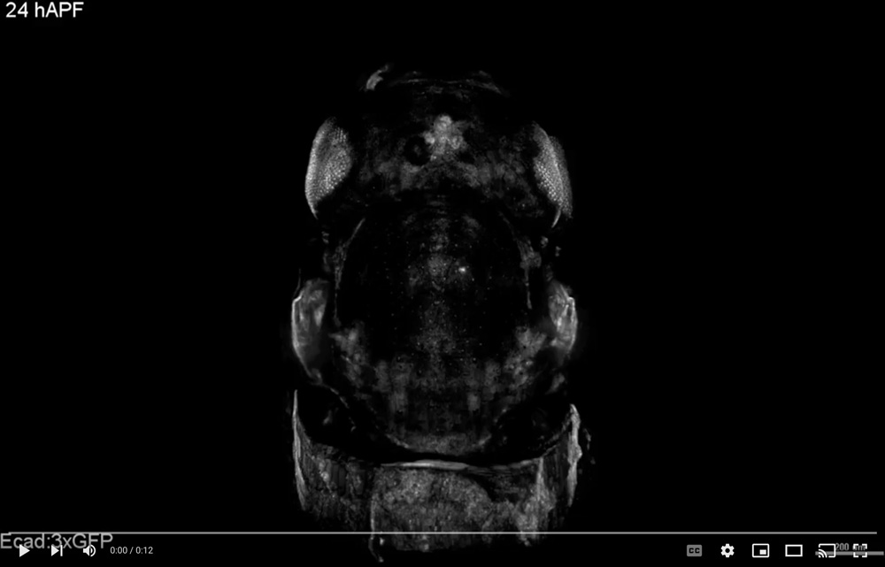
To understand development both cell- and tissue-scale images need to be acquired in three-dimension (3D). Using the Multi-View confocal microScopy (MuViScopy) one can reconstruct the 3D cell and tissue organization of the Drosophila pupa. The movie is an animation of a 3D reconstruction after fusion of images acquired from four angles (0°, 90°, 180° and 270°) by the MuViScope of an Ecad:3xGFP Drosophila pupa at 24 hAPF with a 40x objective to achieve cellular resolution. At each angle, 25 tiles (5×5) were acquired to cover the full dissected part of the animal. The animation starts with a dorsal view of the notum and the head, followed by a zoom cellular resolution on the notum and finishes by a 360° rotation around the A-P axis. Scale bar: indicated on the film according to the zoom factor (in μm).
Video Credit: Oliver Leroy, Eric van Leen, Olivier Renaud
Leroy O, van Leen E, Girard P, Villedieu A, Hubert C, Bosveld F, Bellaïche Y, Renaud O.
Multi-view confocal microscopy enables multiple organ and whole organism live-imaging.
Development 2022 Feb 15; 149(4), dev199760.
