2025 Drosophila Image Award
Winner – Video
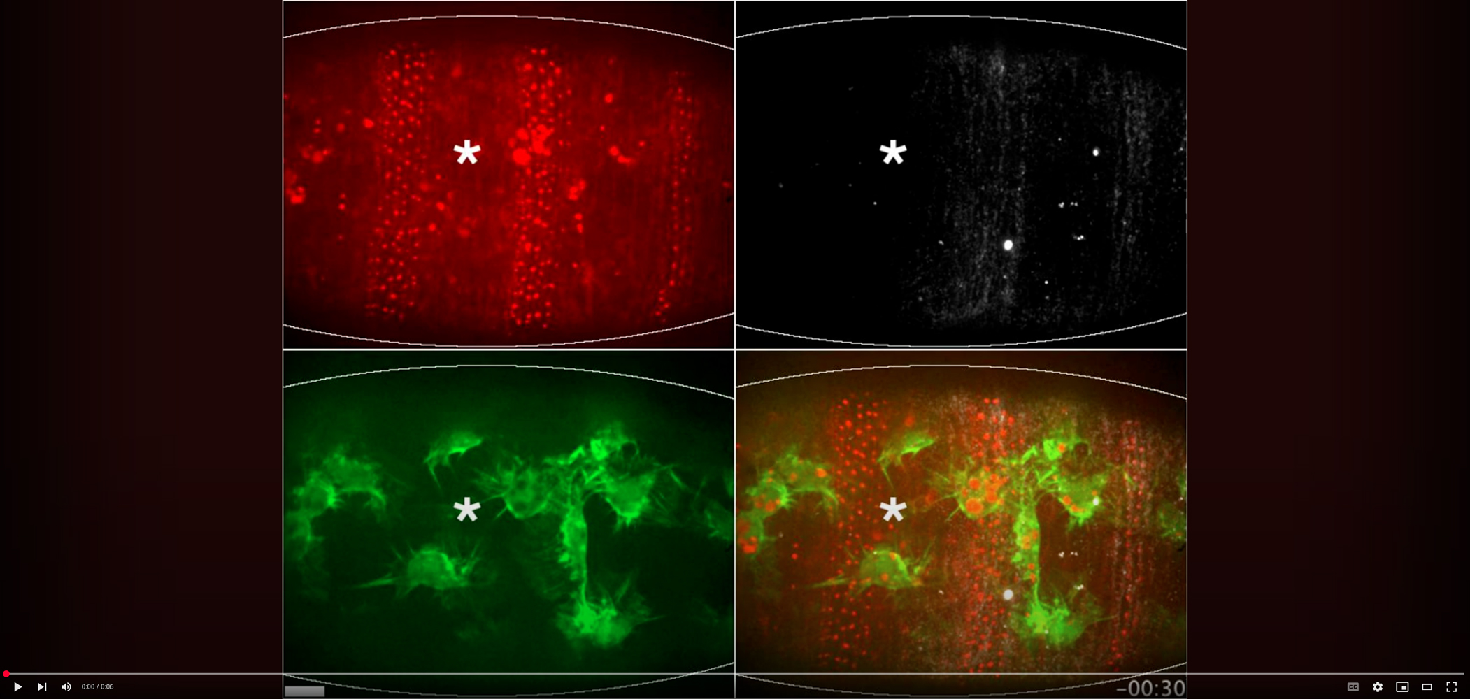
Ring of Fire: three-colour, live-imaging reveals ring of distinct necrosis during wounding of the Drosophila embryo
Three-colour live-imaging of a laser-wounded Drosophila embryo (outlined), with the epithelium labelled with Moesin-mCherry (red), embryonic plasmatocytes (‘macrophages’) labelled with lifeact-GFP (green) and a distinct form of necrosis ringing the edge of the wound labelled with far-red Annexin V (white). Laser-ablation (indicated by asterisk) triggers wounding of the epithelium and the inflammatory recruitment of plasmatocytes. This novel, three colour, live-imaging allowed visualisation of a distinct subset of necrosis, extruded chaotically from the edge of the wound. These necrotic cells are extremely swollen, presenting a unique challenge to the plasmatocytes tasked with clearing this tissue damage. Scale bar = 10 µm, Time = minutes:seconds.
Video Credit: Andrew J. Davidson
Andrew J. Davidson, Rosalind Heron, Jyotirekha Das, Michael Overholtzer and Will Wood
Ferroptosis-like cell death promotes and prolongs inflammation in Drosophila.
Davidson et al., 2024, Nature Cell Biology, 26:1535–1544
http://10.1038/s41556-024-01450-7
Winner – Still Image
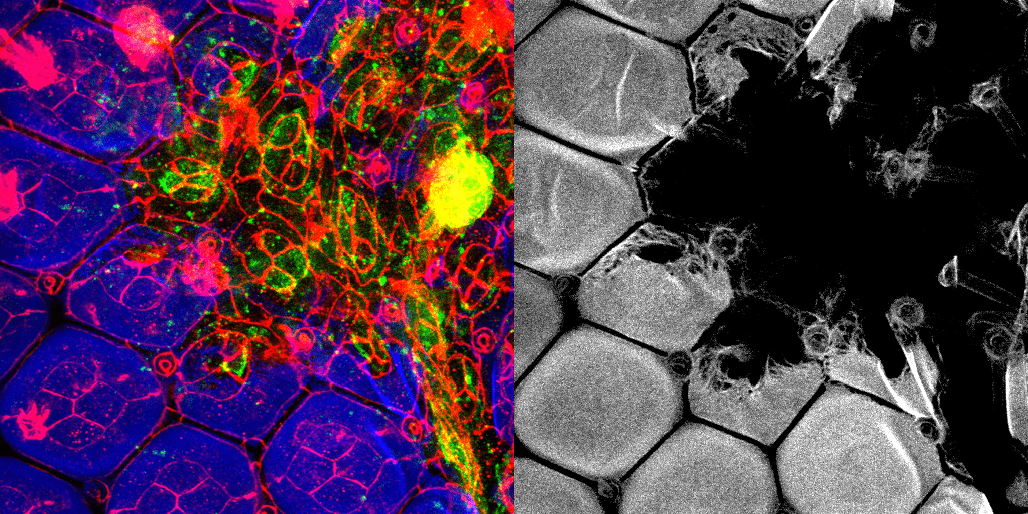
The ZP protein Dusky-like is crucial for apical ommatidial chitin accumulation
The Drosophila corneal lens is an apical extracellular matrix structure composed primarily of a lamellar array of chitin fibers. Our recent study identified a role of the Zona Pellucida domain-containing protein Dusky-like (Dyl) in controlling corneal lens shape. The accompanying image shows ommatidial cell junctions (Arm, red) in a mid-pupal retina containing clones mutant for dyl (GFP, green). We observed that the dyl mutant clones display a complete loss of apical chitin accumulation (Chitin binding domain probe, blue and in single channel) compared to their adjacent wild-type counterparts which produce large amounts of chitin. Additionally, we observe that dyl mutant regions show apical constriction in contrast to the adjoining wild-type ommatidia. The image was created by maximum-intensity z projections obtained using a Leica SP8 II confocal microscope.
Image Credit: Neha Ghosh
Neha Ghosh and Jessica E. Treisman
Apical cell expansion maintained by Dusky-like establishes a scaffold for corneal lens morphogenesis.
Ghosh and Treisman, 2024, Science advances, 10(34), eado4167.
https://doi.org/10.1126/sciadv.ado4167
1st Runner Up – Video
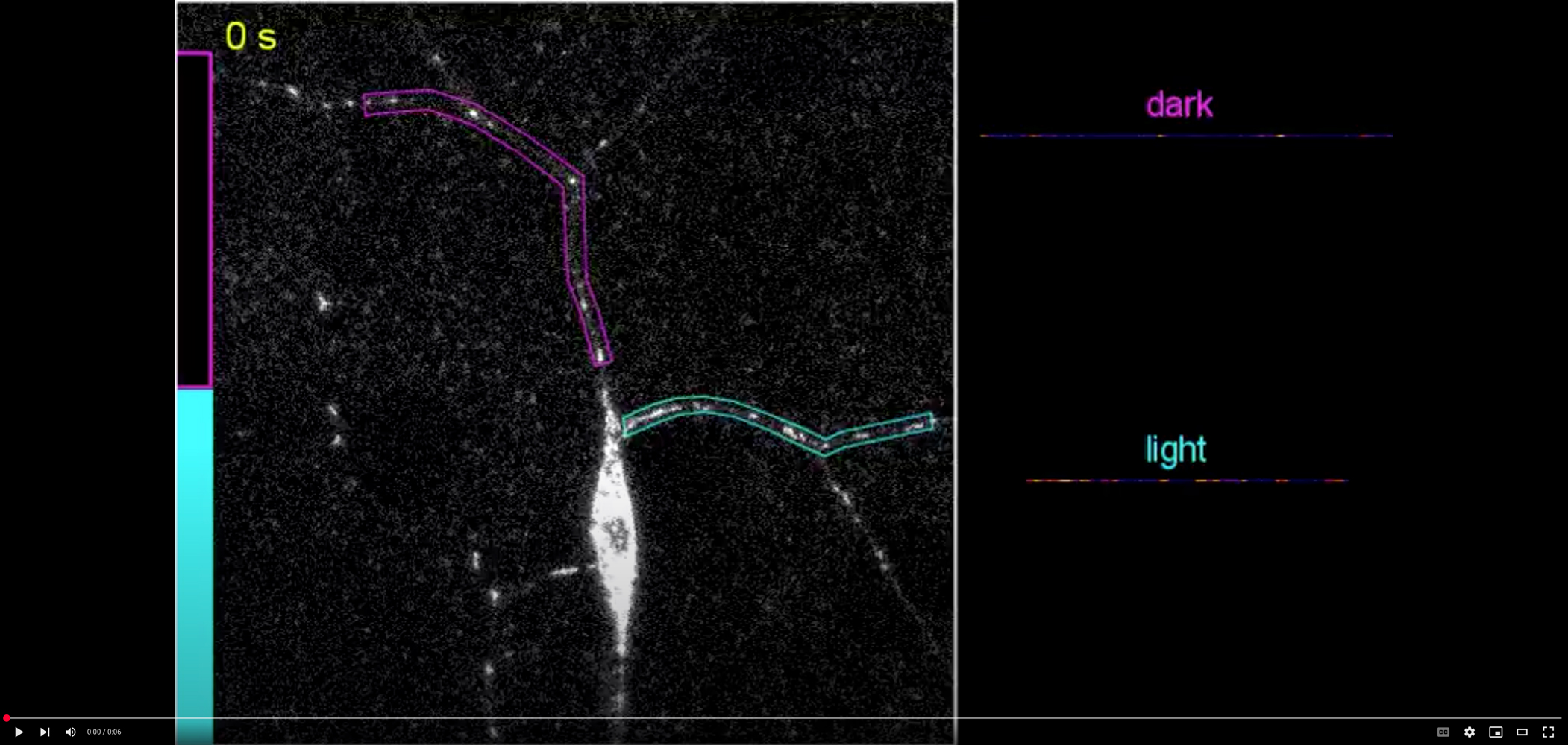
Instant and local trapping of mitochondria by light-induced kinesin-1 inhibition in neuronal dendrites
Kinesin is a microtubule-based motor that transports mitochondria bidirectionally along neuronal dendrites. In this class IV da (C4da) neuron, mitochondria were labeled by mito-mCherry and imaged in a time-lapse experiment. The neuron also expressed OptoTrap to enable trapping and inhibition of kinesin-1 upon blue light illumination. The bottom half of the neuron was exposed to blue laser light (indicated by the blue bar on the left) from the beginning of this movie, while the top half of the neuron was kept in dark (indicated by the black bar). In the illuminated branches, mitochondria showed little or no motility, while mitochondria in the dark region continued moving in both anterograde and retrograde directions. The motility of mitochondria in the outlined dendrite branches (magenta for dark and blue for light) was also visualized in kymographs (right), in which moving mitochondria produce diagonal lines while static mitochondria generate vertical lines. The striking difference in mitochondrial motility between the illuminated and dark regions highlights the power of OptoTrap for manipulating the activity of endogenous proteins with high spatiotemporal precision.
Video Credit: Yineng Xu and Chun Han
Yineng Xu, Bei Wang, Inle Bush, Harriet Aj Saunders, Jill Wildonger and Chun Han
In vivo optogenetic manipulations of endogenous proteins reveal spatiotemporal roles of microtubule and kinesin in dendrite patterning.
Xu et al., 2024, Science Advances, Aug 30;10(35):eadp0138
https://doi.org/10.1126/sciadv.adp0138
1st Runner Up – Still Image
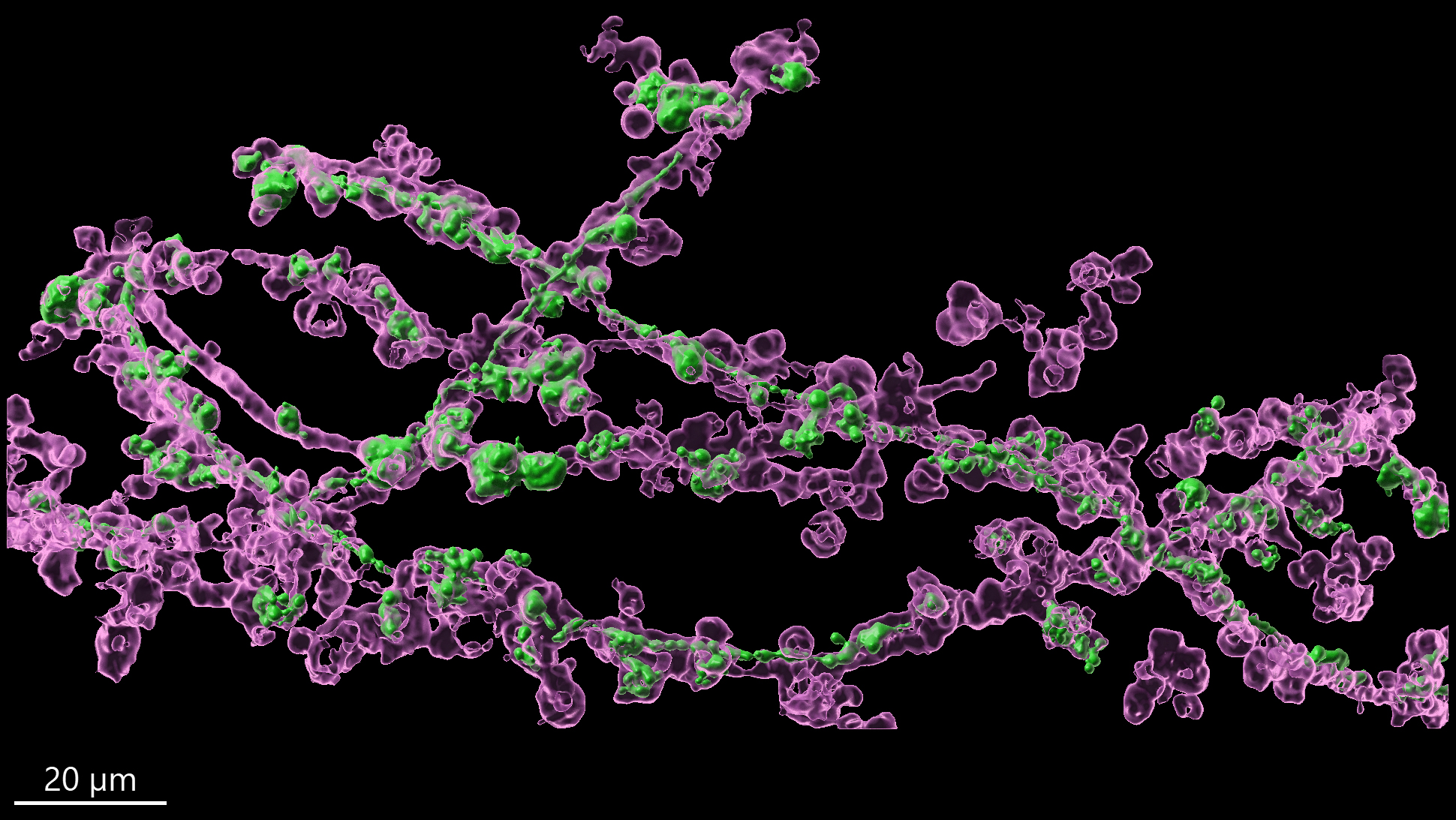
Egg multivesicular bodies elicit an LC3-associated phagocytosis-like pathway to degrade paternal mitochondria after fertilization
Mitochondria are typically maternally inherited, but the mechanisms for eliminating paternal mitochondria after fertilization are not well understood. In our study, we demonstrate that specialized egg-derived multivesicular bodies in Drosophila facilitate the degradation of paternal mitochondria by activating an LC3-associated phagocytosis (LAP)-like pathway, which is generally used by cells to combat invading microbes. The accompanying image shows sperm (paternal) mitochondria (in green) inside an early fertilized egg, prepared for expansion microscopy. The image shows that upon fertilization, these vesicles – marked by Rubicon, a key protein in the LAP pathway (in magenta) – form extensive sheaths around the 2 mm long sperm mitochondrial derivative, promoting its degradation.
Image Credit: Alina Kolpakova and Sharon Ben-Hur
Sharon Ben-Hur, Shoshana Sernik, Sara Afar, Alina Kolpakova, Yoav Politi, Liron Gal, Anat Florentin, Ofra Golani, Ehud Sivan, Nili Dezorella, David Morgenstern, Shmuel Pietrokovski, Eyal Schejter, Keren Yacobi-Sharon and Eli Arama
Egg multivesicular bodies elicit an LC3-associated phagocytosis-like pathway to degrade paternal mitochondria after fertilization.
Ben-Hur et al., 2024, Nature Communications, Jul 8;15(1):5715
https://doi.org/10.1093/genetics/iyad106
2nd Runner Up – Video
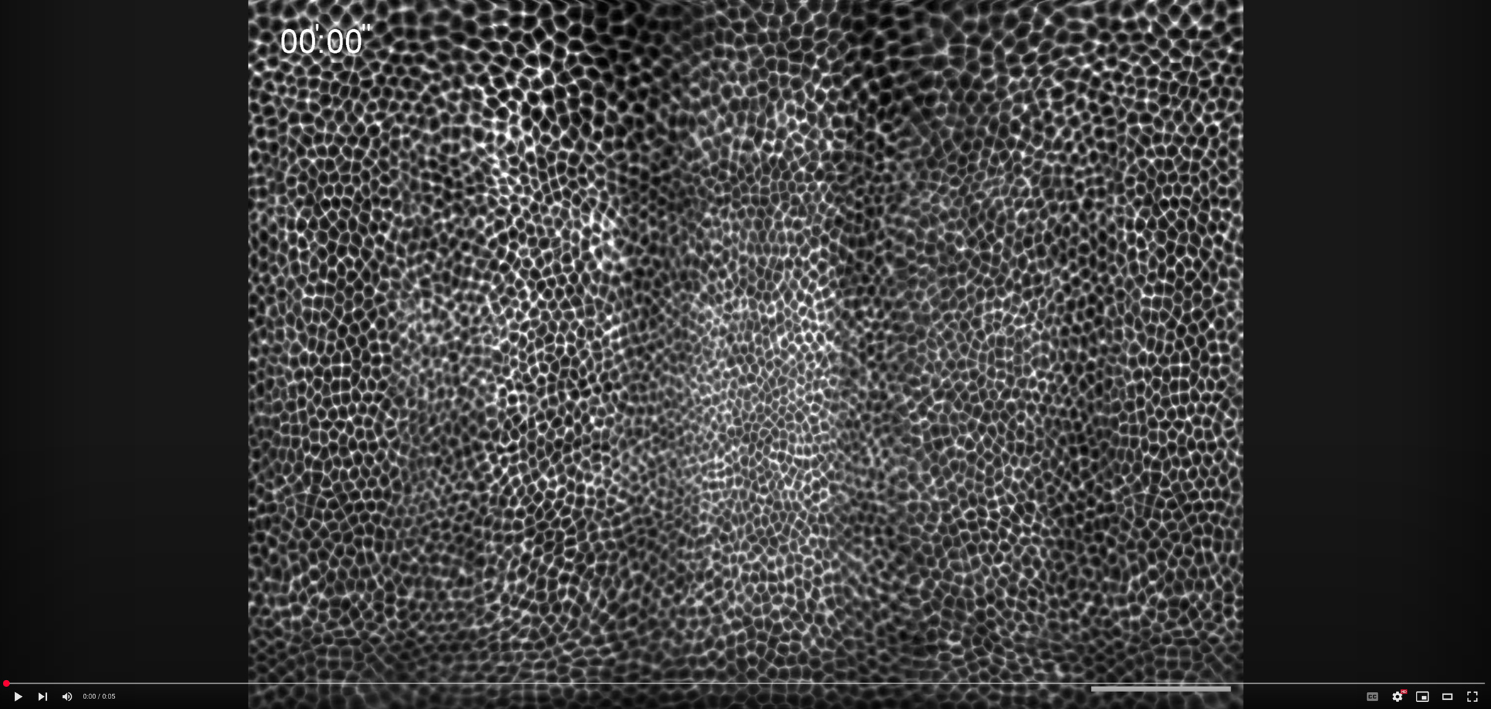
Mercator projection of a living Drosophila embryo during gastrulation when morphogenetic waves sculpt folds and furrows in the blastoderm epithelium
This video shows the formation of the cephalic furrow (see horizontal fold) that separates the head from the trunk region of the embryo. The cephalic furrow results from two folds that bidirectionally propagate and meet on the dorsal side (shown in the center). While the propagation of the ventral furrow (see vertical fold shown twice on the right and left sides) results from embryo scale mechanical forces (Fierling et al. 2022), Popkova and colleagues show that the cephalic furrow is sculpted by a morphogenetic trigger wave (Popkova et al. 2024). The wave is initiated by a group of cells working as a pacemaker, and it propagates along a line under the control of a multidimensional genetic guide.
Anterior (top), posterior (bottom), dorsal (center) and ventral (left and right). Scale bar 50 µm.
Video Credit: Anna Popkova and Matteo Rauzi
Anna Popkova, Urška Andrenšek, Sophie Pagnotta, Primož Ziherl, Matej Krajnc and Matteo Rauzi.
A mechanical wave travels along a genetic guide to drive the formation of an epithelial furrow during Drosophila gastrulation.
Popkova et al., 2024, Developmental Cell Feb 5;59(3):400-414.e5
http://10.1016/j.devcel.2023.12.016
2nd Runner Up – Still Image
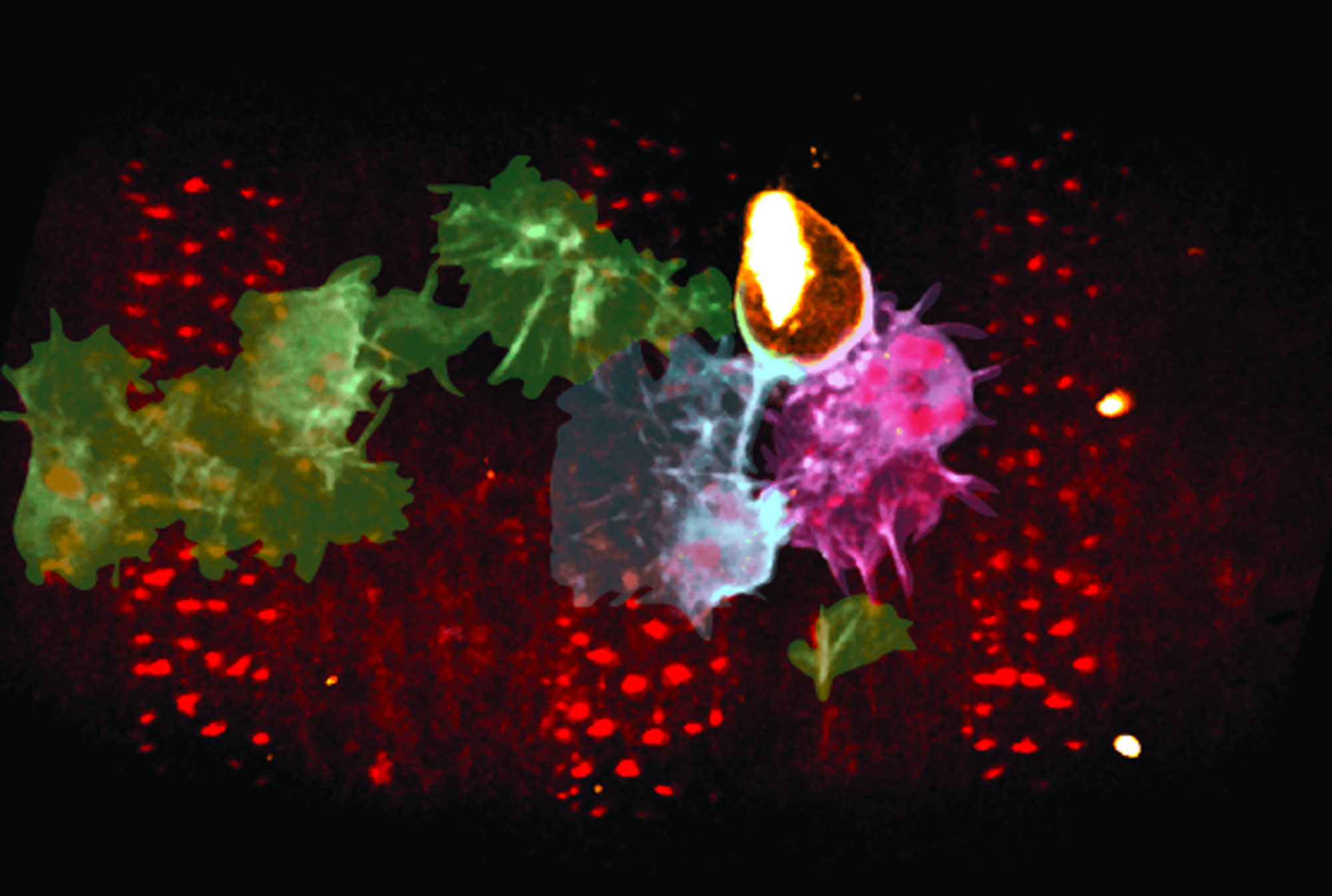
Candy Crush: Embryonic plasmatocytes vying to envelop a necrotic cell
Three-colour live-imaging of a Drosophila embryo, with the epithelium labelled with Moesin-mCherry (red). A single epithelial cell has been irradiated with UV, triggering necrosis, extrusion and labelling with injected far-red Annexin V (orange). Two rapidly recruited, gfp-labelled embryonic plasmatocytes (‘macrophages’, pseudocoloured cyan and magenta, other, uninvolved plasmatocytes, green) wrestle with each other over the swollen necrotic corpse, with neither able to envelope it fully. In the majority of cases, such tussles lead to bursting of the necrotic corpse and the spilling of intracellular material. This novel live-imaging allowed visualisation of necrosis in vivo as well as the challenge it presents to the immune cells tasked with clearing this inflammatory cell death.
Image Credit: Andrew J. Davidson
Andrew J. Davidson, Rosalind Heron, Jyotirekha Das, Michael Overholtzer and Will Wood
Ferroptosis-like cell death promotes and prolongs inflammation in Drosophila.
Davidson et al., 2024, Nature Cell Biology, Sep;26(9):1535-1544
https://doi: 10.1038/s41556-024-01450-7
Honorable Mention – Video
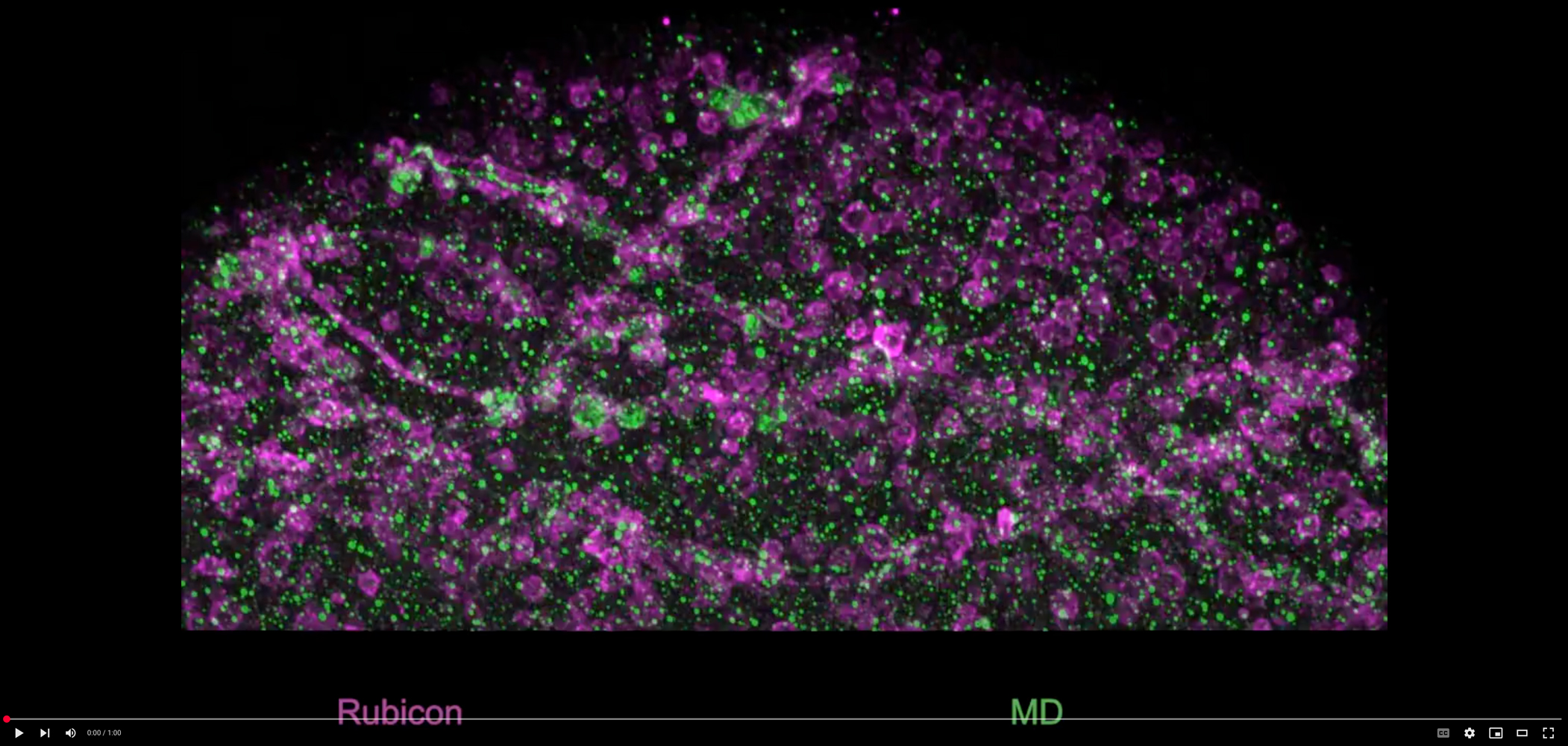
Egg multivesicular bodies elicit an LC3-associated phagocytosis-like pathway to degrade paternal mitochondria after fertilization
Mitochondria are typically maternally inherited, but the mechanisms for eliminating paternal mitochondria after fertilization are not well understood. In our study, we demonstrate that specialized egg-derived multivesicular bodies in Drosophila facilitate the degradation of paternal mitochondria by activating an LC3-associated phagocytosis (LAP)-like pathway, which is generally used by cells to combat invading microbes. The accompanying video shows sperm (paternal) mitochondria (in green) inside an early fertilized egg, prepared for expansion microscopy. The video shows that upon fertilization, these vesicles – marked by Rubicon, a key protein in the LAP pathway (in magenta) – form extensive sheaths around the 2 mm long sperm mitochondrial derivative, promoting its degradation.
Video Credit: Alina Kolpakova and Sharon Ben-Hur
Sharon Ben-Hur, Shoshana Sernik, Sara Afar, Alina Kolpakova, Yoav Politi, Liron Gal, Anat Florentin, Ofra Golani, Ehud Sivan, Nili Dezorella, David Morgenstern, Shmuel Pietrokovski, Eyal Schejter, Keren Yacobi-Sharon and Eli Arama
Egg multivesicular bodies elicit an LC3-associated phagocytosis-like pathway to degrade paternal mitochondria after fertilization.
Ben-Hur et al., 2024, Nature Communications, Jul 8;15(1):5715
https://doi.org/10.1093/genetics/iyad106
Honorable Mention – Still Image
DNA damage responses in the germline
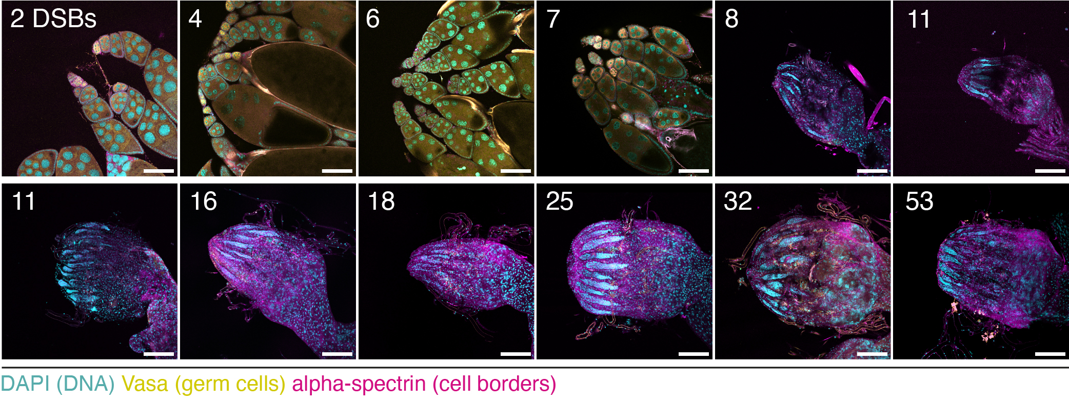
Germ cells carry genetic information across generations. Mechanisms that safeguard genome integrity in the germline, for example by curbing the activity of selfish genetic elements like transposons, are paramount to ensure the faithful transmission of genetic material. Yet how germ cells respond to genome damage has not been systematically investigated. The panel shows images of Drosophila melanogaster ovaries, with germ cells labelled by Vasa (yellow). In these germ cells, different dosages of DNA double strand breaks (DSBs) were induced by targeting the Cas9 endonuclease to a specific number of (non-coding) genomic sites. When few sites were targeted (i.e. DSB dosage was low), ovary morphology resembled the wild-type. Targeting more than 7-8 sites resulted in rudimentary ovaries completely devoid of germ cells, indicating that germ cells are sensitive to DSBs in a dosage-dependent manner. These findings establish DNA damage tolerance thresholds as a crucial protective mechanism for maintaining genome integrity and germ cell survival during development.
Image Credit: Gloria Jansen
Gloria Jansen, Daniel Gebert, Tharini Ravindra Kumar, Emily Simmons, Sarah Murphy and Felipe Karam Teixeira
Tolerance thresholds underlie responses to DNA damage during germline development.
Jansen et al., 2024, Genes & Development, 38(13-14), 631-654.
https://doi.org/10.1101/gad.351701.124
Honorable Mention – Video
Deep neural networks automatically tag high-speed Drosophila cardiac walls and assess contractile dynamics
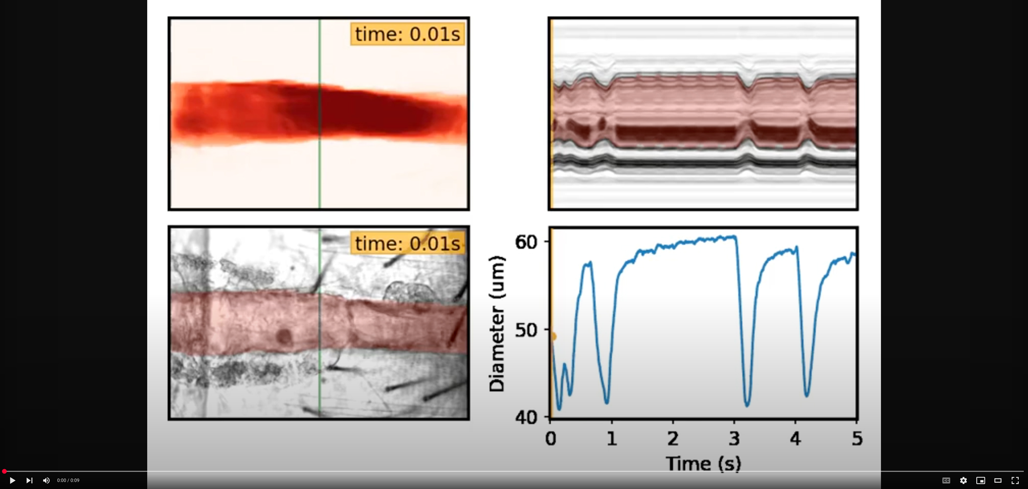
Analysis of high-speed Drosophila cardiac video reveals important details about contractile dynamics, aging, and cardiovascular disease. However, the task remains highly manual and tedious. We demonstrate how deep neural networks (DNN) can be applied for the automated tracking of Drosophila cardiac walls. The figure shows that, with DNN-based tracking, full video beating patterns (M-Mode) and contractility can be recovered with little human effort. The demonstrated approach is applied for large-scale analysis of aging and cardiac dysfunction models.
Video Credit: Yash Melkani and Aniket Pant
Aniket Pant, Yash Melkani, Yiming Guo, Girish Melkani
Automated assessment of cardiac dynamics in aging and dilated cardiomyopathy Drosophila models using machine learning.
Pant et al., 2024, Communications Biology, Jun 7;7(1):702
https://doi.org/10.1038/s42003-024-06371-7
Honorable Mention – Still Image
Third instar larval brain as a model for virus-host interactions
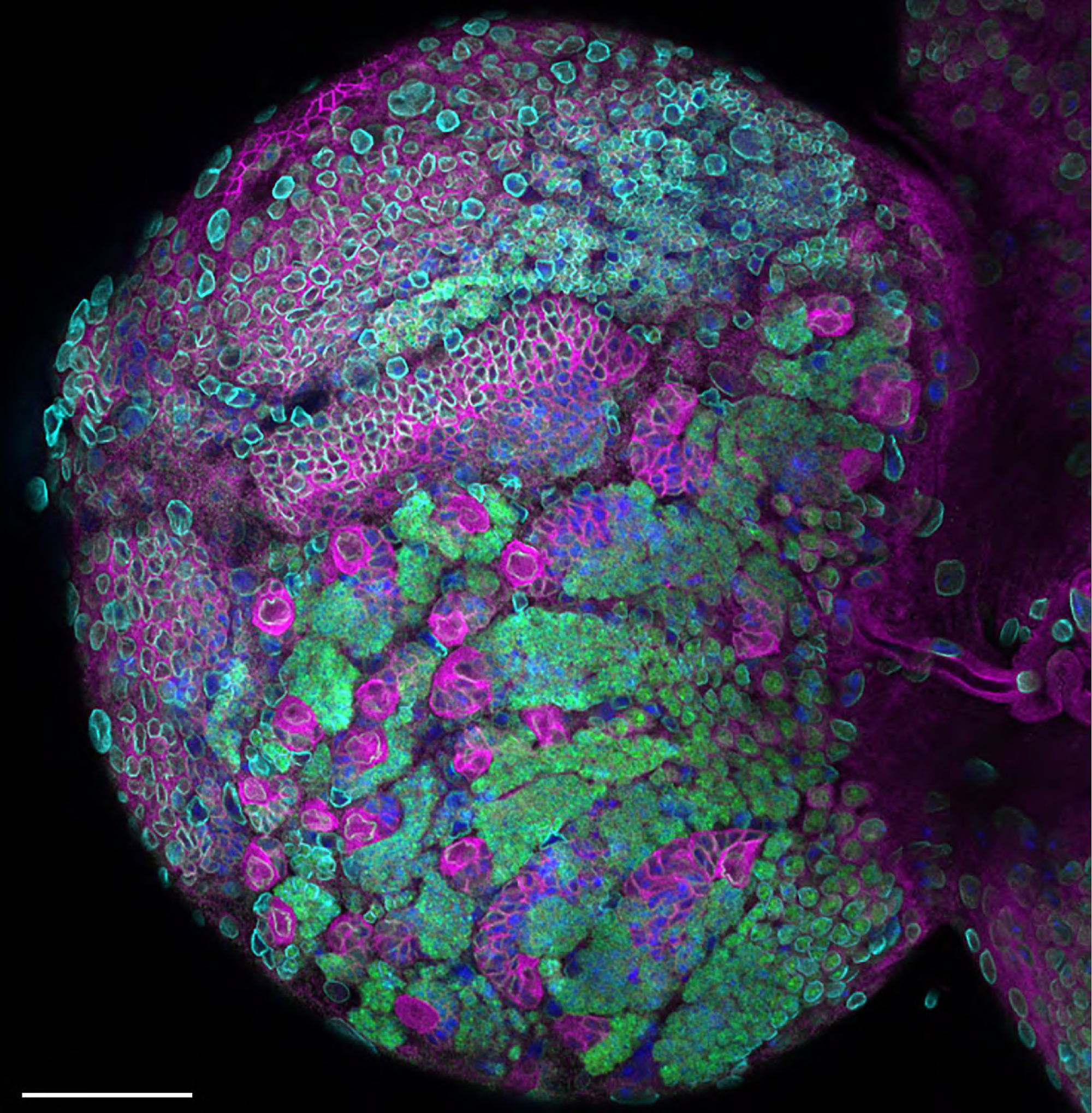
Drosophila third instar larval brain stained for aPKC (magenta), Elav (green, neurons), Lamin (cyan, nuclear envelope), DNA was stained using DAPI (blue). The large magenta cells are neural stem cells, whereas the smaller green cells are differentiating neuronal lineages produced by an asymmetrically dividing stem cell. The image shows a partial z-projection.
Image Credit: Nichole Link
Nichole Link, J. Michael Harnish, Brooke Hull, Shelley Gibson, Miranda Dietze ,Uchechukwu E. Mgbike, Silvia Medina-Balcazar, Priya S. Shah and Shinya Yamamoto
A Zika virus protein expression screen in Drosophila to investigate targeted host pathways during development.
Link et al., 2024, Disease Models and Mechanisms, 17 (2): dmm050297.
https://doi.org/10.1242/dmm.050297
