2024 Drosophila Image Award
Winner – Video
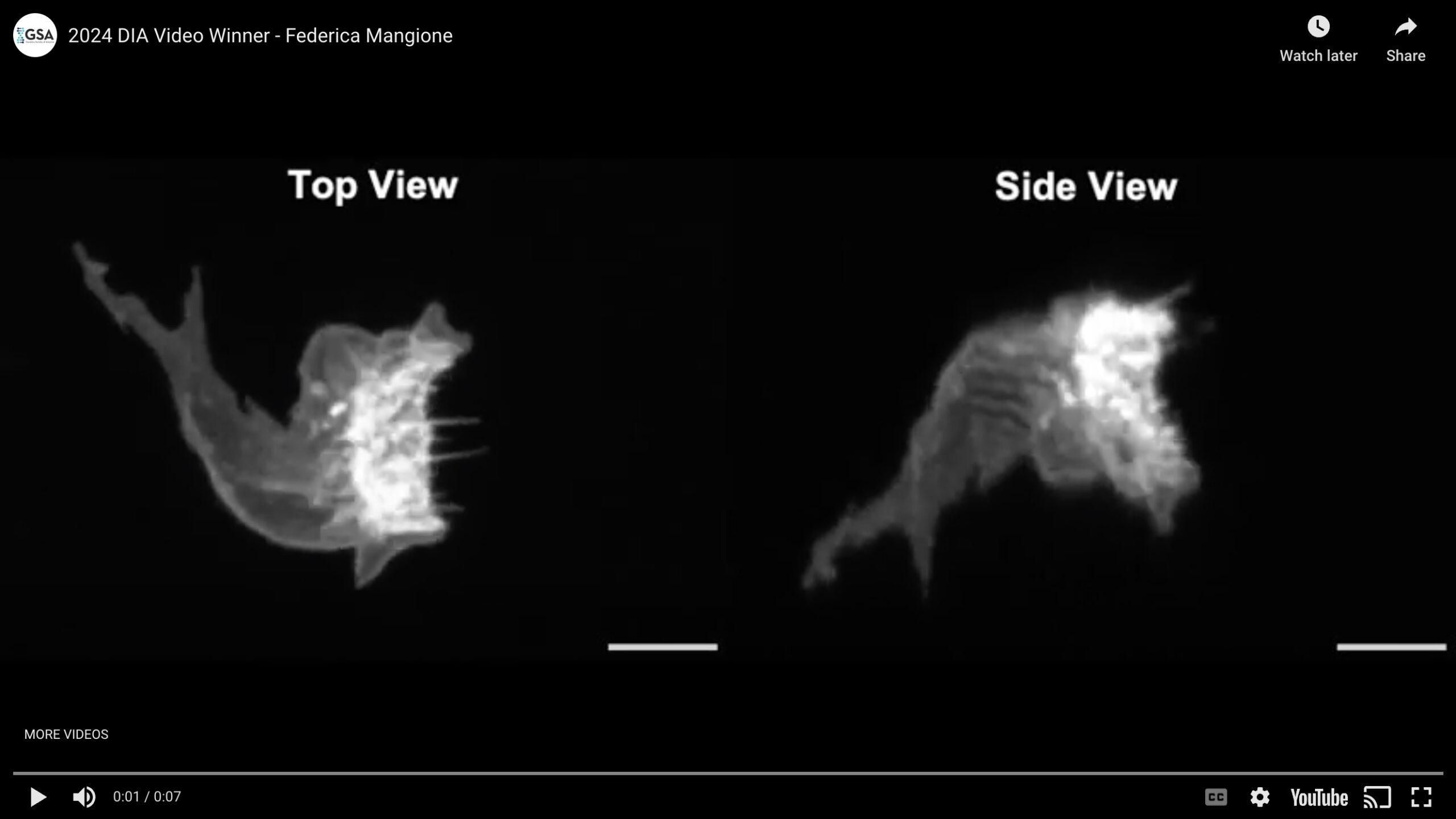
Morphological Differentiation of the F-Cell, a Specialized Epidermal Cell Embracing the Tactile Bristle
Specialized cellular structures endow the epidermis with the sense touch. While characterizing the terminal differentiation of the tactile bristles, the most abundant sensory organs within the Drosophila epidermis, we uncovered the association of a specialized epidermal cell, the F-Cell, by mid pupal stage. We found that F-Cells are recruited by the differentiating bristles from the surrounding epidermis to complete the assembly of the tactile bristle, enabling touch sensation in adulthood. The F-Cell and its differentiation were visualized for the first time in vivo by live imaging with confocal microscopy. This video captures the morphological changes of the F-Cell as it associates with a neighbouring bristle. Between 58 hours and 68 hours after puparium formation (hAPF), the morphology of the F-Cell changes markedly, culminating in a ring-like shape as it fully embraces the bristle by 68 hAPF. This unique shape distinguishes the F-Cell from other epidermal cells in Drosophila. For this video, the morphology of the F-Cell was made visible by the expression of the membrane marker myr::GFP under the control of 25c01-GAL4. Displayed scale bars are 5 µm. Our findings provide insight into the cellular organization underlying touch sensation and its developmental basis.
Video Credit: Federica Mangione
Mangione F, Titlow J, Maclachlan C, Gho M, Davis I, Collinson L, Tapon N.
Co-option of epidermal cells enables touch sensing.
Nat Cell Biol. 2023 Apr;25(4):540-549.
Winner – Still Image
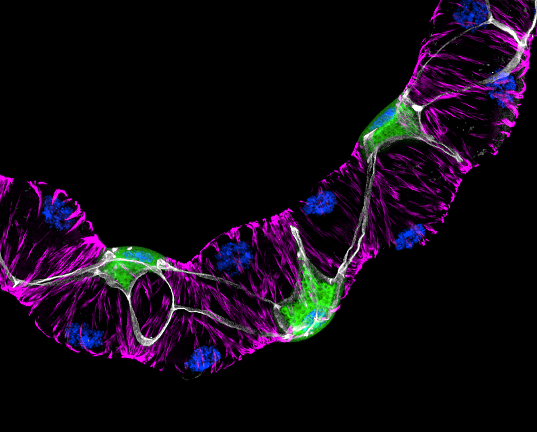
Cellular organisation of the fly’s kidney
Detail of an adult Drosophila Malpighian (renal) tubule expressing membrane-bound GFP (green) in stellate cells – a population secondary intercalated cells required for transport of water – under the direction of c724GAL4, with staining of nuclei (DAPI, blue) and F-actin microfilaments (TRITC-conjugated phalloidin, magenta) in principal cells and stellate cells. Septate junctions are stained using an antibody for the junctional component Discs-large (anti-Dlg1, grey). The image was created by tiling of three separate maximum-intensity z projections obtained using a Nikon AX R NSPARC confocal.
Image Credit: Anthony Dornan
Dornan AJ, Halberg KV, Beuter LK, Davies SA, and JAT Dow.
Compromised junctional integrity phenocopies age-dependent renal dysfunction in Drosophila Snakeskin mutants.
J Cell Sci. 136(19) (2023). doi: 10.1242/jcs.261118
1st Runner Up – Video
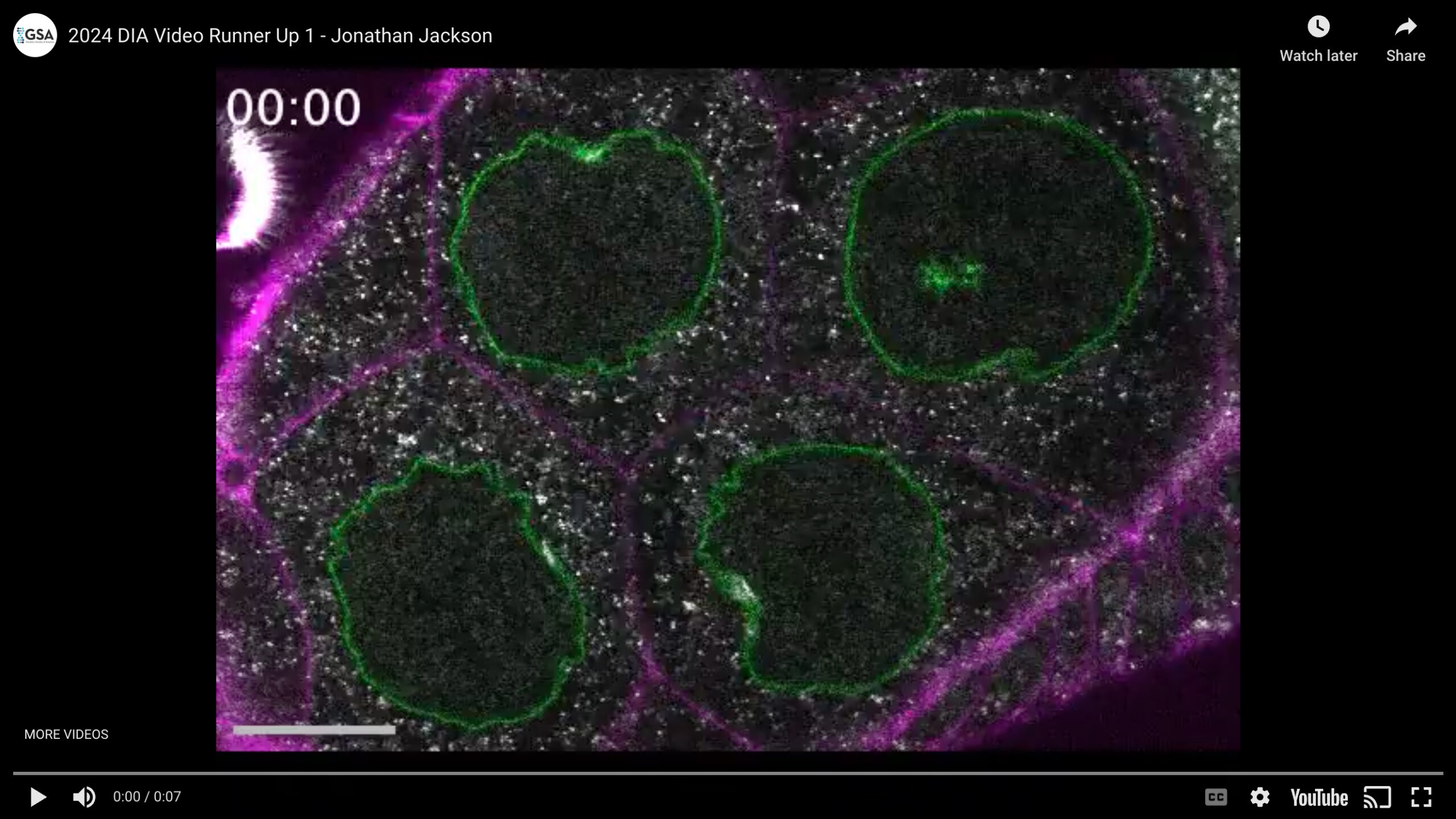
Fluctuations of the nuclear envelope in Drosophila nurse cells are associated with cytoplasmic movement
Drosophila oocytes develop inside a structure known as the egg chamber, connected to fifteen support germline cells known as nurse cells. Fluctuating wrinkles appear in the nuclear envelopes of the nurse cells as development proceeds. In this video, fluctuations of cytoplasmic contents (gray) can be seen associated with deformations of the nuclear envelope (green), with the cell membranes shown in magenta. Scale bars are 20 µm; green is Nup107, a nuclear pore complex protein; magenta is the CellMask plasma membrane dye, and gray is label-free signal acquired using confocal reflectance microscopy.
Video Credit: Jonathan Jackson
Jackson JA*, Romeo N*, Mietke A, Burns KJ, Totz JF, Martin AC, Dunkel J, Imran Alsous J. *(equal contribution)
Scaling behaviour and control of nuclear wrinkling.
Nat. Phys. 2023 Sep 18;19:1927-1935.
1st Runner Up – Still Image
Identification of a non-canonical phosphate-sensing organelle in the Drosophila midgut
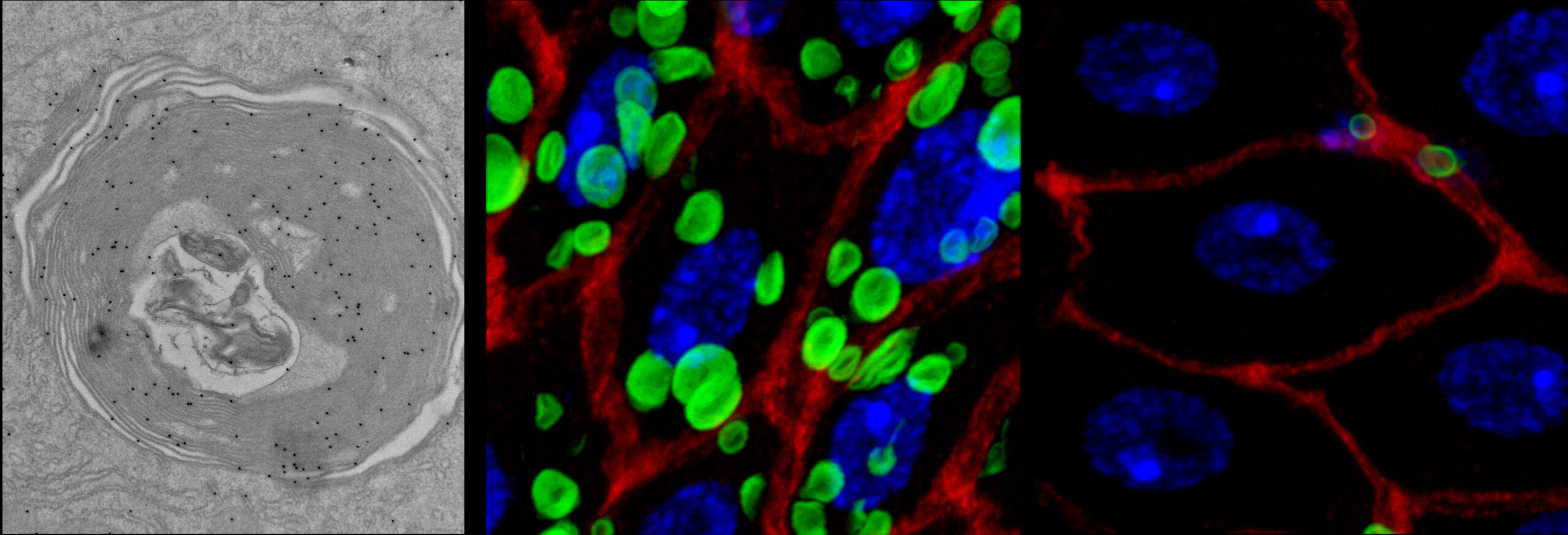
CG10483 (PXo), a Drosophila ortholog of inorganic phosphate transporter, is the membrane marker of a previously uncharacterized organelle (PXo body) in the fly intestinal epithelium. The electron micrograph (left) captures the multilamellar morphology of a PXo body, with examples of nanogold labeling for GFP-PXo highlighted by red arrowheads. PXo bodies function as an intracellular phosphate reservoir in the enterocytes. Their size and density are sensitive to the availability of dietary inorganic phosphate. As the confocal images (right) unveiled, PXo bodies underwent degradation when flies were fed with phosphonoformic acid (PFA), a potent inhibitor of phosphate uptake. The fluorescent stainings: green (GFP-PXo), red (Discs large, labeling enterocyte cell borders), blue (DAPI, labeling nuclei).
Image Credit: Chiwei (Charles) Xu
Chiwei Xu*+, Jun Xu*, Hong-Wen Tang, Maria Ericsson, Jui-Hsia Weng, Jonathan DiRusso, Yanhui Hu, Wenzhe Ma, John M. Asara, Norbert Perrimon+. *co-first; +co-correspondence
A phosphate-sensing organelle regulates phosphate and tissue homeostasis.
Nature, 2023 May;617(7962):798-806; PMID: 37138087.
2nd Runner Up – Video
The Fusion Pore of Large Secretory Vesicles Facilitates a Distinct Mode of Exocytosis – “Membrane Crumpling”
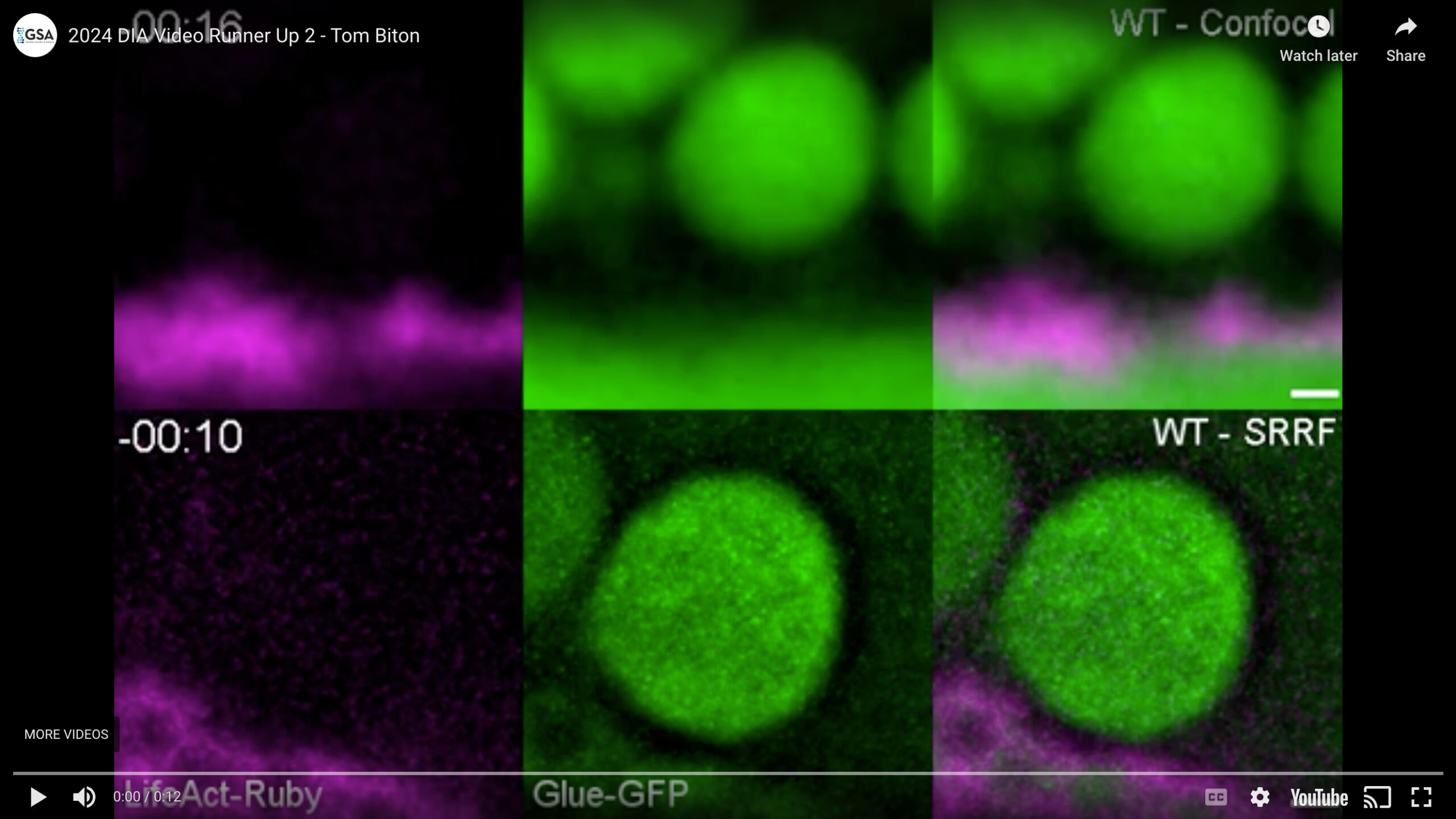
The large secretory vesicle (LSV) exocytic fusion pore expands, stabilizes at a wide diameter, and eventually constricts. This facilitates actomyosin-mediated content release and membrane crumpling while preventing full collapse and vesicle integration. Membrane crumpling ultimately functions as a mechanism to maintain apical membrane homeostasis in a secretory tissues like the Drosophila salivary gland. Conventional confocal imaging (Confocal; top panels) allows to follow secretion dynamics from fusion (00:00; visible by ‘vesicle swelling’; content marker ‘Glue-GFP’; Green), through actomyosin recruitment (00:32-00:48; actin marker ‘LifeAct-Ruby’ as a proxy; Magenta), and finally actomyosin mediated content release (01:04-03:12). To characterize pore dynamics, we visualize it using live super-resolution radial fluctuations imaging (SRRF; bottom panels). The pore expands for ~30 sec following fusion (00:00-00:30), then stabilizes at an expanded diameter while actomyosin assembles on the LSV (00:30-01:00). The pore eventually constricts in parallel to actomyosin compression of the LSV, which results in content release and membrane crumpling (01:00-05:20). Both panels are maximum intensity projections of an image stack. Scale bars = 1 µm.
Video Credit: Tom Biton
Tom Biton, Nadav Scher, Shari Carmon, Yael Elbaz-Alon, Eyal D.Schejter, Ben-Zion Shilo, and Ori Avinoam.
Fusion pore dynamics of large secretory vesicles define a distinct mechanism of exocytosis.
J Cell Biol (2023) 222 (11): e202302112.
2nd Runner Up – Still Image
Getting in Touch with the F-Cell
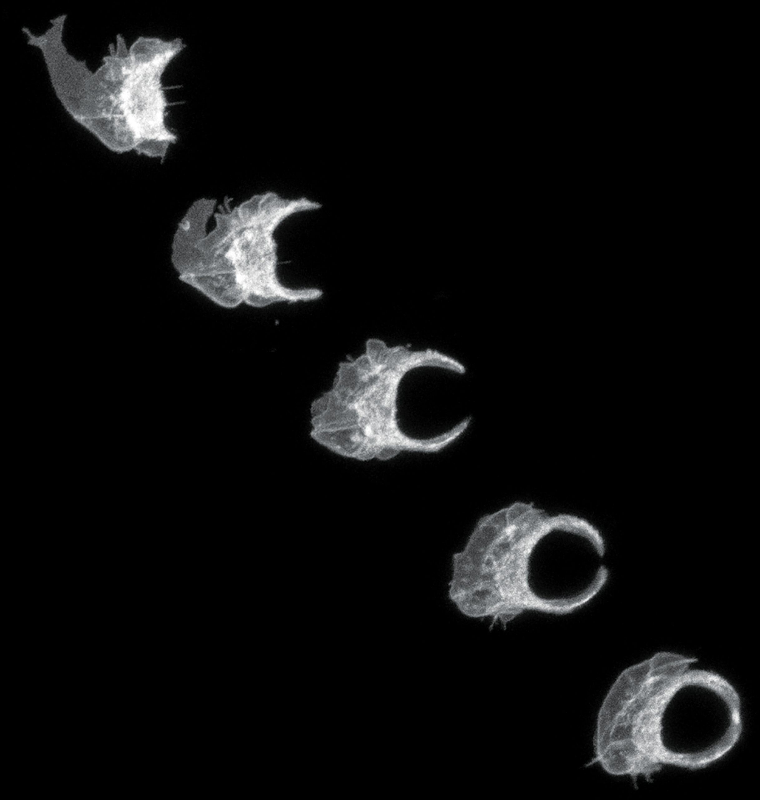
Specialized cellular structures endow the epidermis with the sense touch. While characterizing the terminal differentiation of the tactile bristles, the most abundant sensory organs within the Drosophila epidermis, we uncovered the association of a specialized epidermal cell, the F-Cell, by mid pupal stage. We found that the F-Cell is co-opted by the differentiating bristles from the surrounding epidermis to complete the assembly of the tactile bristle, enabling touch sensation in adulthood. The F-Cell and its differentiation were visualized for the first time in vivo by live imaging with confocal microscopy. This image displays a time course of the shape changes underlying the morphological differentiation of the F-Cell between 58 hours and 68 hours after puparium formation. Note the marked shape changes in the F-Cell, the only epidermal cell that remodel its shape to embrace a neighbouring bristle. Here, the morphology of the F-Cell encircling the tactile bristle was made visible by the expression of the membrane marker myr::GFP under the control of 25c01-GAL4. Our findings revealed a crucial role for epidermal cells during the morphogenesis of the sensory system of touch.
Image Credit: Federica Mangione
Mangione F, Titlow J, Maclachlan C, Gho M, Davis I, Collinson L, Tapon N.
Co-option of epidermal cells enables touch sensing.
Nat Cell Biol. 2023 Apr;25(4):540-549.
Honorable Mention – Video
Acute manipulation and real-time visualization of exocytosis
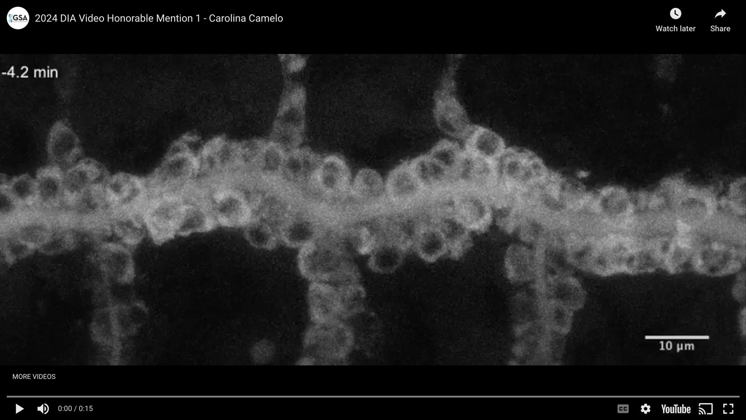
Intracellular trafficking and exocytosis of the secreted protein Serpentine (Serp) was synchronized and visualized in the tracheal system of a Drosophila embryo by means of the Retention Using Selective Hooks (RUSH) system. Exit of Serp-GFP from the endoplasmic reticulum was triggered at t=0 min by injection of biotin into the embryo.
Video Credit: Carolina Camelo
Glashauser, J.*, Camelo, C.*, Hollmann, M., Backer, W., Jacobs, T., Isasti Sanchez, J., Schleutker, R., Förster, D., Berns, N., Riechmann, V., and Luschnig, S. (2023).
Acute manipulation and real-time visualization of membrane trafficking and exocytosis in Drosophila.
Developmental Cell 58(8): 709-723.e7.
Honorable Mention – Still Image
Glial wrapping provides insulation to the axons and also limits their access to the extracellular ion reservoir
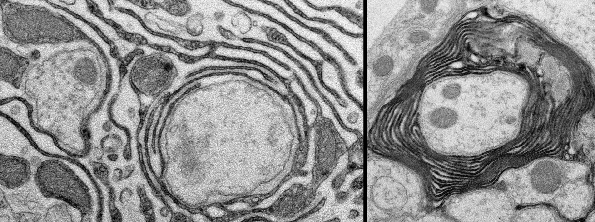
The formation of glial lacunae around the proximal segments of motoaxons, where voltage-gated sodium channels accumulate, enlarges the extracellular ion pool (left). A collapse of the lacunar spaces distally to regions with high density of voltage-gated ion channels results in the formation of myelin-like structures (right).
Image Credit: Simone Rey
Simone Rey, Henrike Ohm, Frederieke Moschref, Dagmar Zeuschner, Marit Praetz, Christian Klämbt.
Glial-dependent clustering of voltage-gated ion channels in Drosophila precedes myelin formation.
eLife
Honorable Mention – Video
Cyclic myosin flows are associated with cardioblast migration
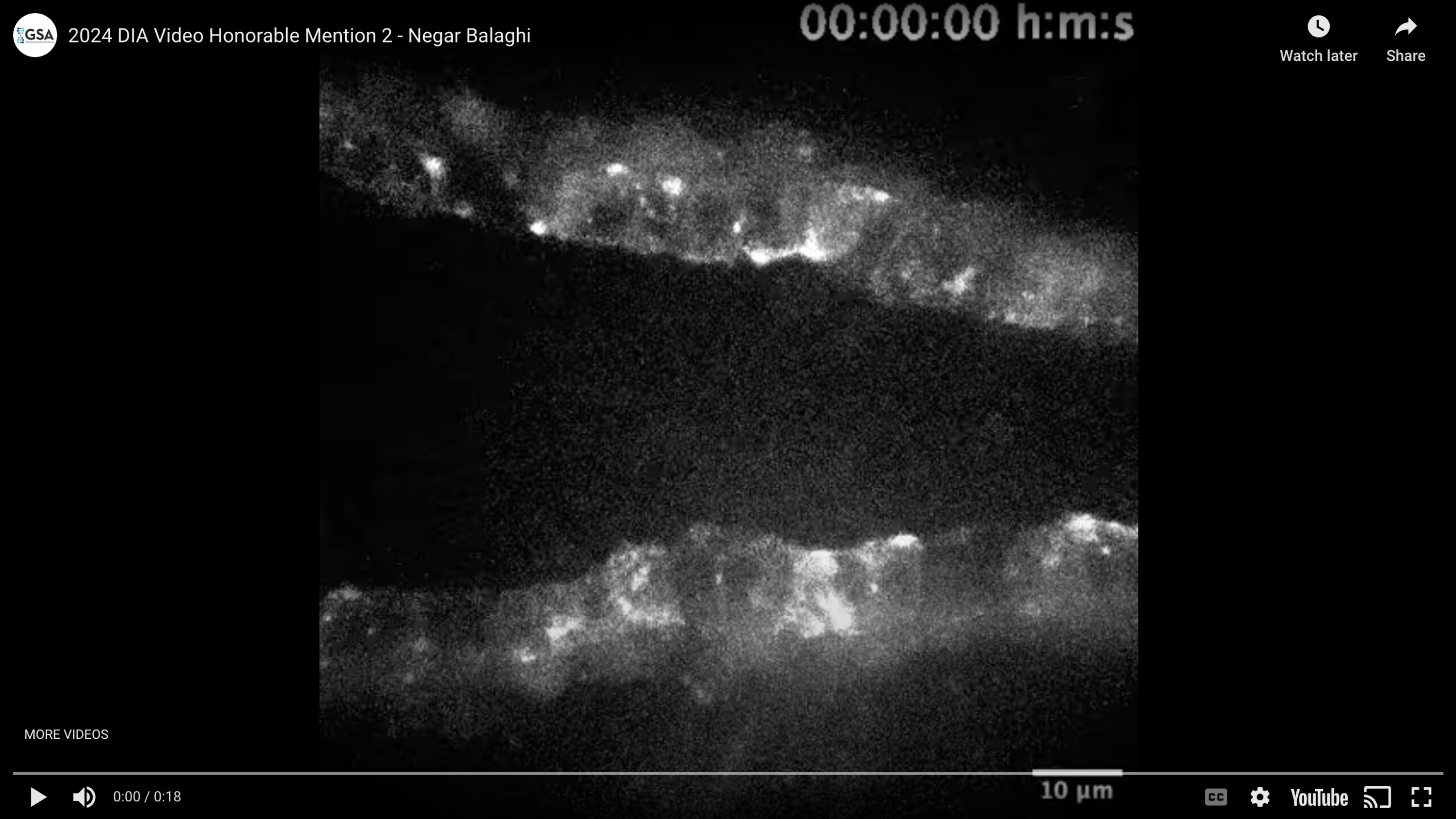
In Drosophila, the embryonic heart is formed by 52 pairs of contralateral cardiac precursors (cardioblasts) that migrate dorsally and medially to form a tube. Cardioblasts migrate through a sequence of forward and backward steps; the forward steps are greater in amplitude and duration, resulting in the net forward movement of the cells. The molecular motor non-muscle myosin II generates mechanical forces for cell movement. We examined myosin dynamics in cardioblasts using time-lapse microscopy of embryos expressing a GFP-tagged form of the myosin light chain (sqh:GFP) specifically in cardiac progenitors. We found that within each cardioblast, myosin displayed an alternating pattern of localization between the leading and trailing ends of the cell. The periodic pattern of myosin localization produced striking oscillatory waves. Myosin waves promoted alternative contraction of the leading and trailing edges of the cardioblasts and periodic cell shape changes. We found that the periodic myosin waves and cell shape changes were coupled with a supracellular actin cable at the trailing edge of the cardioblasts. The actin cable acts as a stiff border that rectifies myosin-induced oscillations into asymmetrical force production for forward cardioblast migration.
Video Credit: Negar Balaghi
Balaghi N, Erdemci-Tandogan G, McFaul CM, Fernandez-Gonzalez R.
Myosin waves and a mechanical asymmetry guide the oscillatory migration of Drosophila cardiac progenitors.
Dev. Cell. 2023 Jul 24;58(14):1299-1313.
Honorable Mention – Still Image
Colorful mosaic of neuroblast lineages encased by their glial niche in the larval central nervous system, upon loss of the adhesion molecule Wrapper
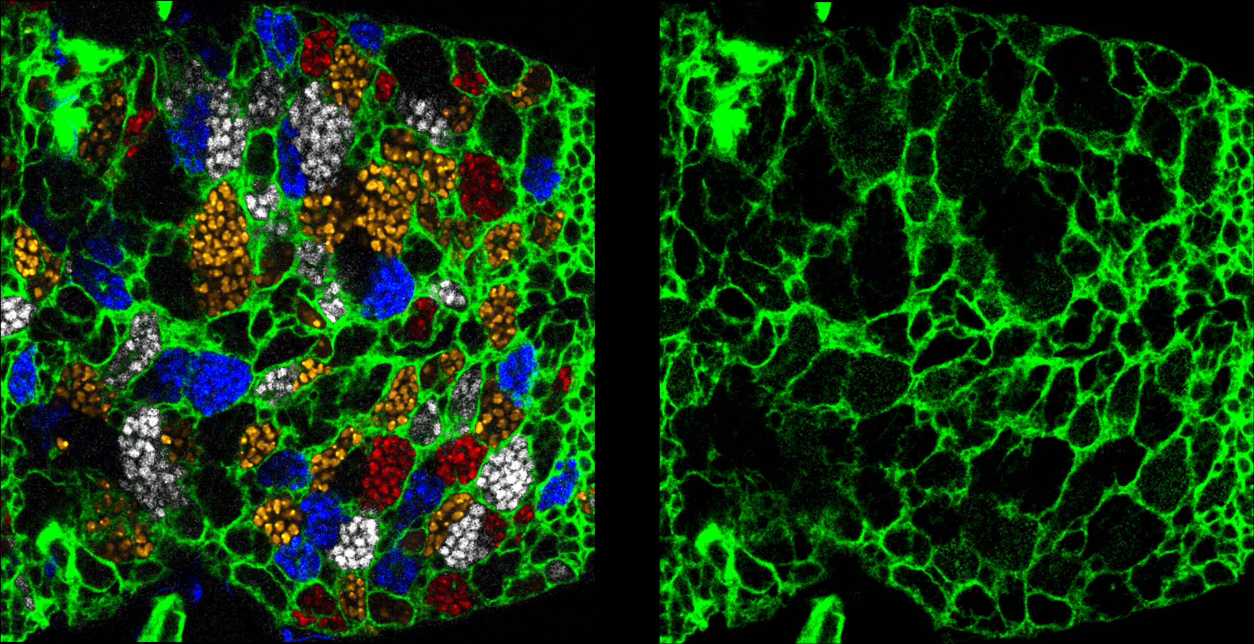
Cortex glia form a membrane network encasing individual neuroblasts and their secondary lineages in a tailored membrane chamber. Stochastic multicolour labelling (nucleus-localised Raeppli-NLS: White, Cyan, Orange and Red) revealed an abnormal organisation when the adhesion molecule Wrapper is lost in the cortex glia (membrane labelled with Nrv2::GFP, green). During normal development, Wrapper binds to Neurexin-IV expressed in the neuroblast lineages. When this interaction is altered, individual encasing is lost, as shown by the existence of large chambers containing several neuroblast lineages grouped together. Strikingly, disruption of Wrapper/Neurexin-IV interaction specifically during development is also associated with locomotor hyperactivity in the adult.
Image Credit: Pauline Spéder
Banach-Latapy A, Rincheval V, Briand D, Guénal I, Spéder P.
Differential adhesion during development establishes individual neural stem cell niches and shapes adult behaviour in Drosophila.
PLoS Biol. 2023 Nov 9;21(11):e3002352.
Honorable Mention – Video
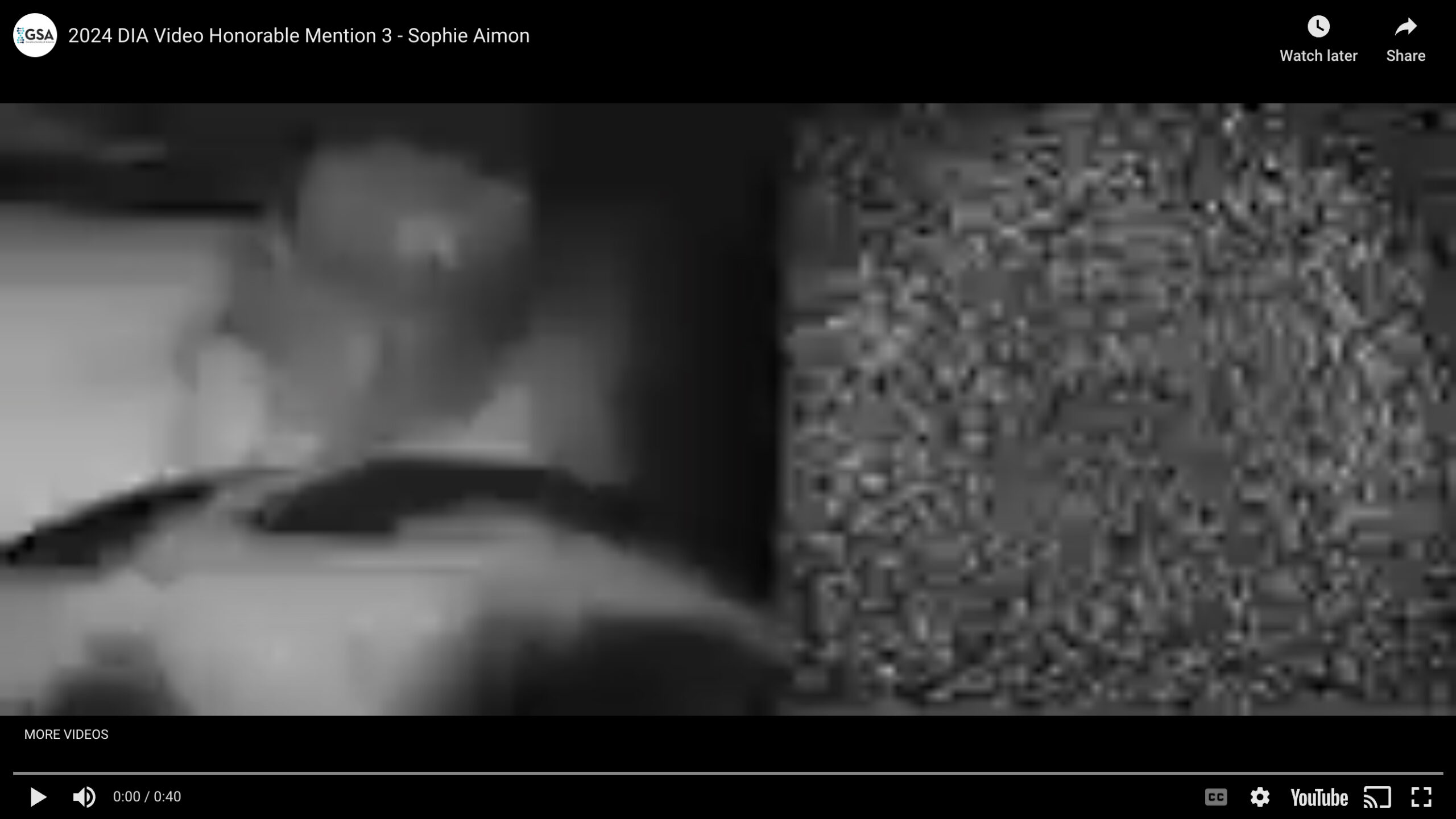
Video showing broad activation of dopamine neurons during walking and turning
The fly genotype is TH-GAL4, DDC-GAL4, UAS-GCaMP6f. Note the specific patterns when the fly turns left or right. These patterns match the expression pattern of PPM2-LW dopaminergic neurons.
Video Credit: Sophie Aimon
Sophie Aimon, Karen Y Cheng, Julijana Gjorgjieva, Ilona C Grunwald Kadow (2023)
Global change in brain state during spontaneous and forced walk in Drosophila is composed of combined activity patterns of different neuron classes.
eLife 12:e85202
Honorable Mention – Still Image
Netrin pathway is required for axon targeting of Drosophila medulla projection neurons in the visual system
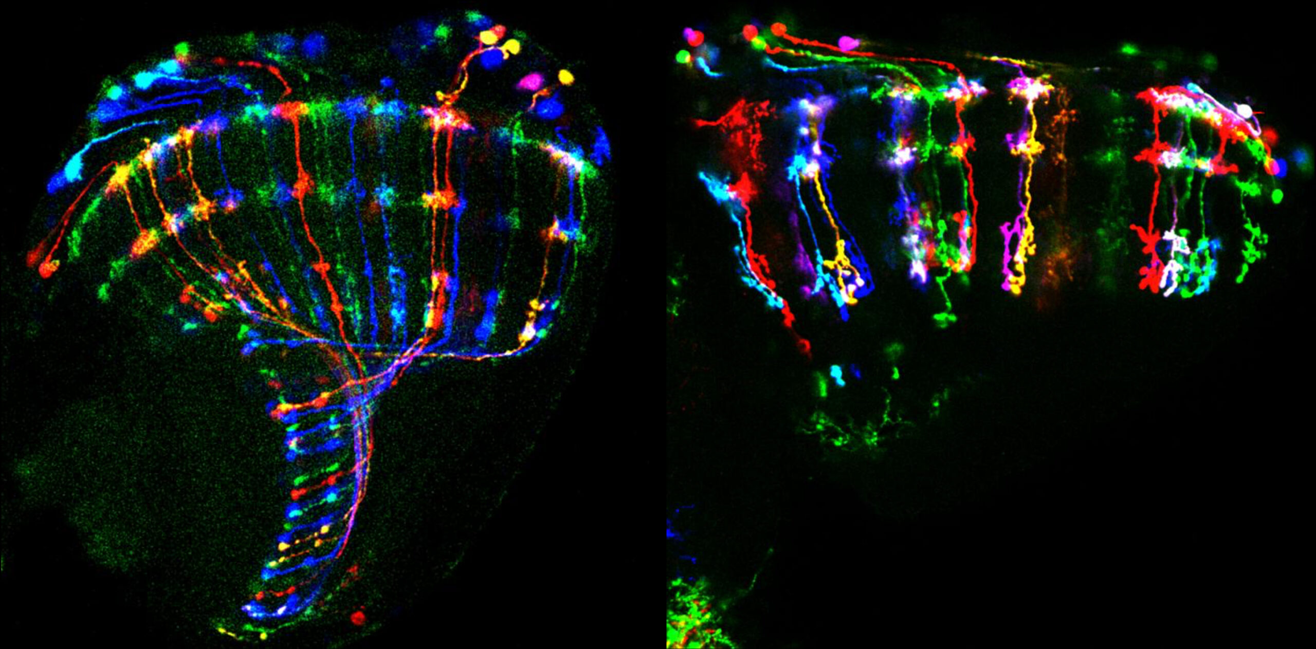
Multicolor FlpOut (MCFO) technique was used to label individual medulla projection neurons, specifically Tm3 neurons. In the wild-type brain (illustrated on the left), the cell bodies of Tm3 neurons are located within the medulla cortex. The dendrites of these neurons extend across various layers of the medulla neuropil, and the axons terminate in the next-order neuropil, the lobula. Conversely, in the Netrin mutant (illustrated on the right), the majority of Tm3 axons fail to reach the lobula neuropil. Instead, these axons exhibit a reversion, redirecting back towards the medulla neuropil. This aberrant axonal pathfinding in the Netrin mutants strongly suggests that the Netrin signaling pathway plays a crucial role in the axonal targeting of Tm3 neurons.
Image Credit: Yu Zhang
Yu Zhang, Scott Lowe, Andrew Z. Ding, Xin Li
Axon targeting of Drosophila medulla projection neurons requires diffusible Netrin and is coordinated with neuroblast temporal patterning.
Cell Reports,Volume 42, Issue 3, 2023.
Honorable Mention – Video
Spatiotemporal remodeling of extracellular matrix orients epithelial sheet folding
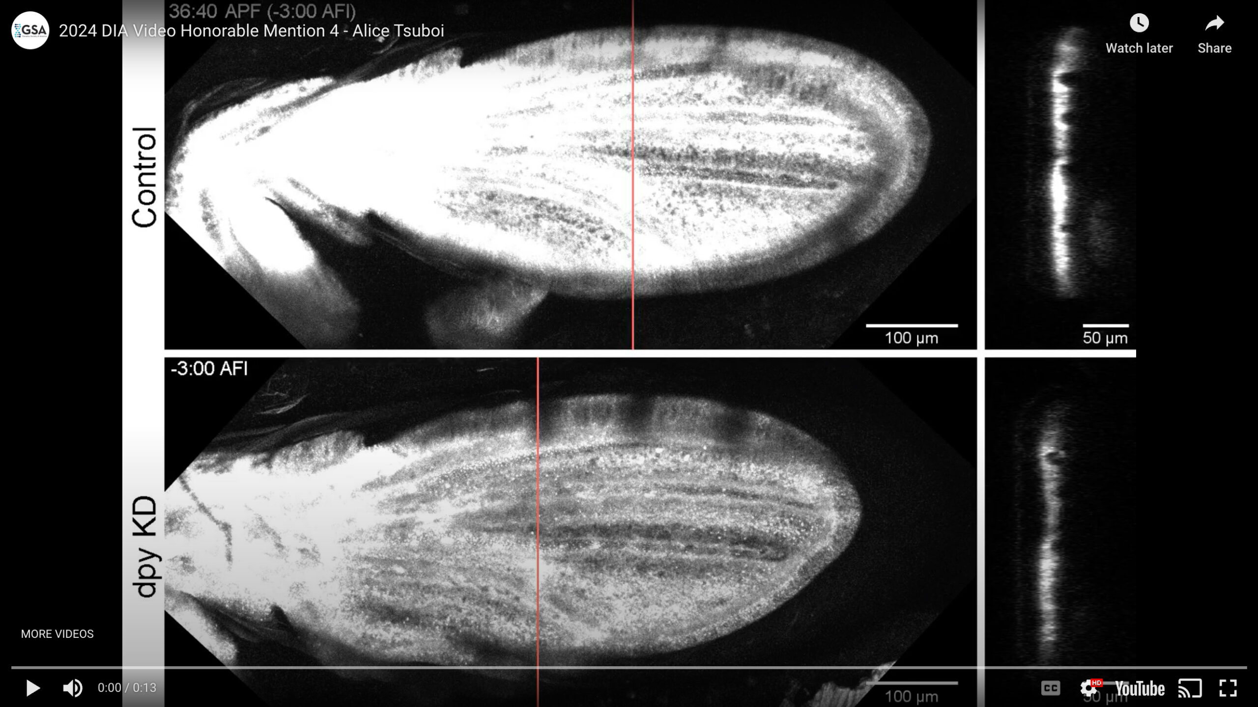
How tissues achieve stereotypic morphology remains an important question in developmental biology. We discovered that apical ECM protein Dumpy (Dpy) specifies the buckling position and direction: Dpy not only covers apical surface of wing tissue, but forms spatially patterned anchorage between wing tissue and its surrounding cuticle, which instructs the buckling pattern along veins. Moreover, after controlling the buckling direction, Dpy undergoes degradation in a specific spatial pattern, which further specifies the positions of the higher-order 3D folding. While ECM has been considered to be a passive mechanical element that follows the primary drivers for tissue shaping (i.e., patterned cellular actomyosin contractility), our work provides wide-ranging implications for the instructive role of patterned ECM remodeling in tissue morphogenesis.
Video Credit: Alice Tsuboi
Alice Tsuboi, Koichi Fujimoto, and Takefumi Kondo.
Spatiotemporal remodeling of extracellular matrix orients epithelial sheet folding.
Science Advances. 2023 Sep;9(35):eadh2154.
