2022 Drosophila Image Award
Winner – Video
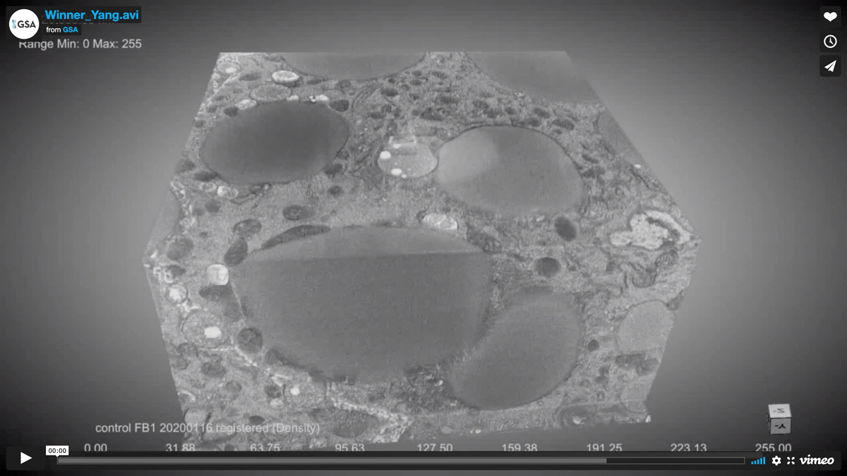
High resolution imaging of the ER-Golgi interface through FIB-SEM
ER exit sites (ERES) of the collagen-producing larval fat body imaged through focused ion beam scanning electron microscopy (FIB-SEM). In each ERES-Golgi unit, the Golgi apparatus (pink) is wrapped by a cup of fenestrated ER sheets (green). Ribosome-devoid ERES regions (grey) are found on the concave side of the cup. Between ERES and Golgi, there are numerous small vesicles (yellow) consistent in size with regular COPII and COPI vesicles. No evidence of larger “megacarrier” vesicles is observed. However, tubular continuities (blue) are seen that directly connect ERES and Golgi. This characterization of ERES-Golgi units provides key insights for understanding secretory transport of large protein cargos at the ER-Golgi interface.
Video Credit: Ke Yang
Yang K, Liu M, Feng Z, Rojas M, Zhou L, Ke H, Pastor-Pareja JC.
ER exit sites in Drosophila display abundant ER-Golgi vesicles and pearled tubes but no megacarriers.
Cell Rep. 2021 Sep 14;36(11):109707
Winner – Still Image
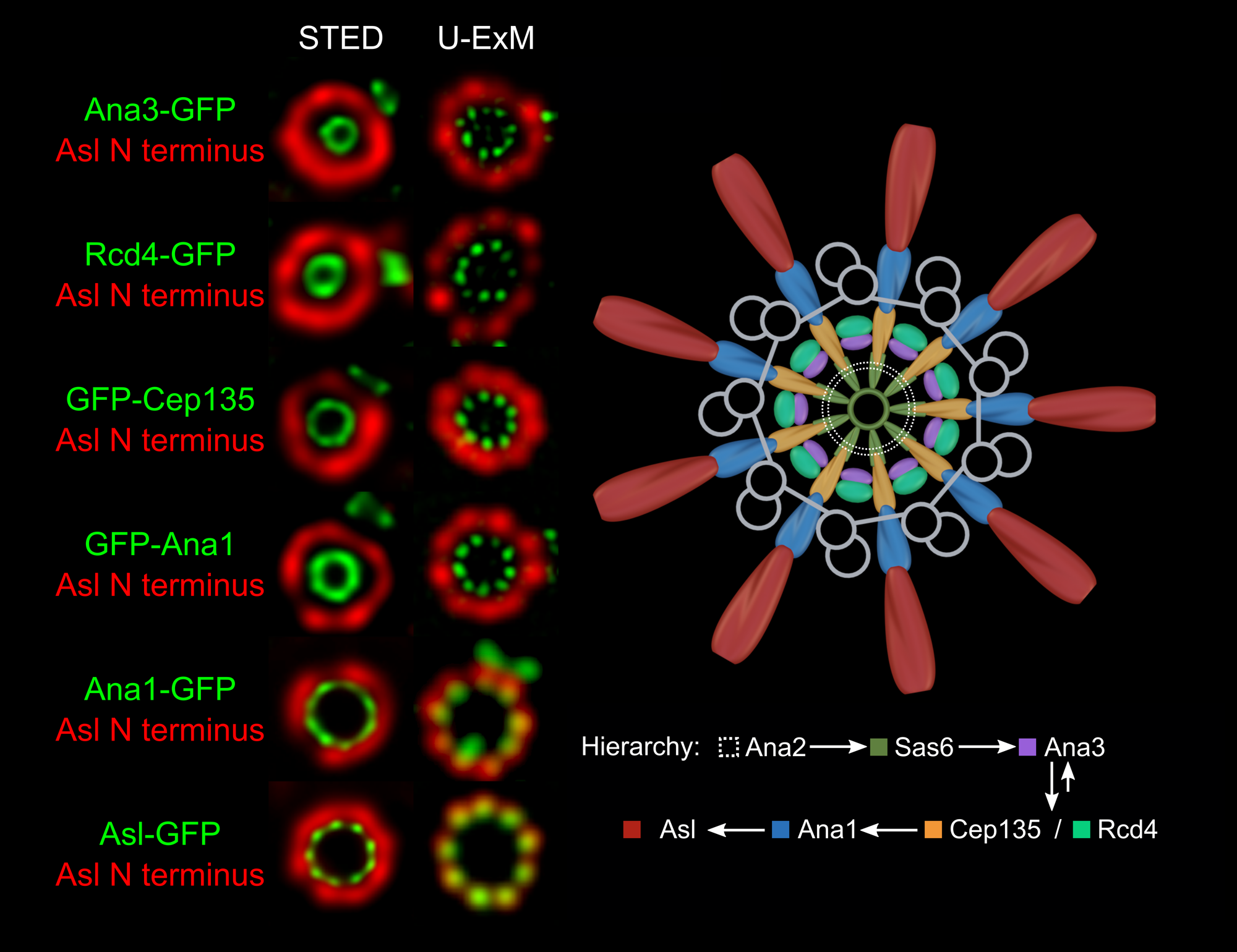
Centrosome Biogenesis under Sub-Diffraction Resolution
For stimula00ted emission depletion (STED) microscopy, Drosophila cultured cell lines constitutively expressing GFP-fused protein were fixed and immunostained with GFP-booster Atto488 (green) and antibody against the N-terminus of Asl (red). For ultra-expansion microscopy (U-ExM), Drosophila cultured cell lines constitutively expressing GFP-fused protein were embedded in polyacrylamide hydrogel. After digestion, expansion, labelling with antibodies against GFP (green) and the N-terminus of Asl (Red), gels were visualized via three dimensional structured illumination microscopy (3D-SIM). Micrograms demonstrated the radial distributions of multiple toroid-shaped proteins. Further analyses revealed relative distribution of pairwise proteins along angular axis. The diameter of each centriolar protein matches its assembly order during centrosome duplication that concerts with cell-division cycle.
Image Credit: Yuan Tian, Yuxuan Yan, Jingyan Fu
Tian Y, Wei C, He J, Yan Y, Pang N, Fang X, Liang X, Fu J.
Superresolution characterization of core centriole architecture.
J Cell Biol. 2021 Apr 5;220(4)
1st Runner Up – Video
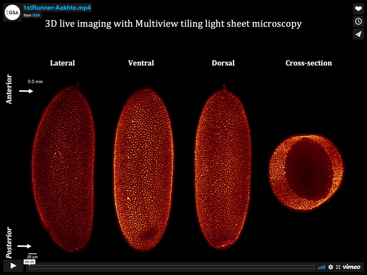
Multiview tiling light sheet microscopy for 3D high-resolution live imaging
The novel high-resolution Multiview tiling light sheet microscope (MT-SPIM) was applied to dynamically image the distribution of MyoII-GFP fluorescence intensities during Drosophila embryo development. The isotropic resolution of MT-SPIM provides entire functional live imaging sequences of known subcellular and supracellular actomyosin arrangements during early gastrulation. The alterations in MyoII-GFP were followed from furrow formation at the cellularization stage, when the MyoII-GFP fluorescence intensities shift radially from apical to basal of the cells. The ventral domain of actomyosin activities are raised locally to contribute to the creation of the ventral furrow, the cephalic furrow and the posterior midgut primordium.
Video Credit: Mostafa Aakhte
Aakhte M, Müller HJ.
Multiview tiling light sheet microscopy for 3D high-resolution live imaging.
Development. 2021 Sep 15;148(18):dev199725
1st Runner Up – Still Image
Nephrocyte endocytosis
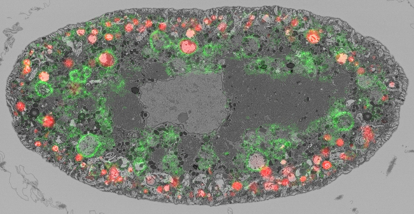
Drosophila melanogaster renal cells, called nephrocytes, filter and endocytose extracellular macromolecules. They function to regulate the composition of the hemolymph and to counteract the harmful effects of nutritional imbalances such as a high fat diet. The image is of a single pericardial nephrocyte obtained with correlative light and electron microscopy (CLEM), showing plasma membrane lacunae, an extensive endoplasmic reticulum surrounding the nucleus, and endosomes labelled with dextran (red) and Rab7::YFP (green).
Image Credit: Aleksandra Lubojemska and Azumi Yoshimura
Lubojemska A, Stefana MI, Sorge S, Bailey AP, Lampe L, Yoshimura A, Burrell A, Collinson L, Gould AP.
Adipose triglyceride lipase protects renal cell endocytosis in a Drosophila dietary model of chronic kidney disease.
PLoS Biol. 2021 May 4;19(5)
2nd Runner Up – Video
Budding Process of the First Egg Chamber
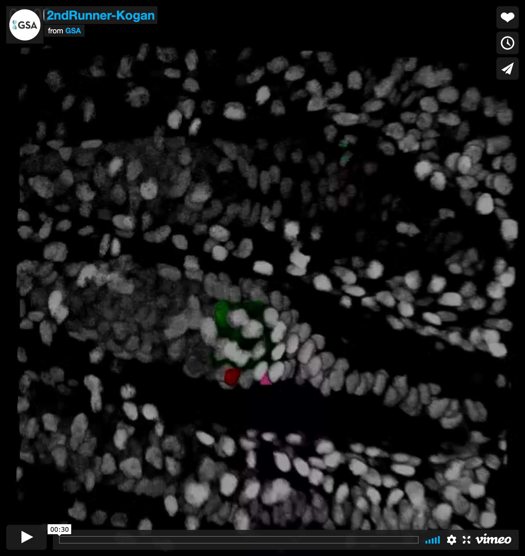
The budding of the first egg-chamber of the Drosophila ovary marks a crucial event in development and cell fate specification. This process was visualized for the first time by live imaging of a developing ubi-GFP labeled ex-vivo pupal ovary. In the pupal germarium, germline cells are interspersed with somatic intermingled cells. We found that an additional pool of somatic cells progressively accumulates posterior to intermingled cells from 12 to 36h after puparium formation (APF). Some of these cells form a multilayered cone or crown around the posterior of the developing germarium (an extra-germarial crown (EGC)); the rest intercalate to form a more posterior “basal stalk”. This video tracks development from 48h APF and shows a germline cyst (green) moving into the EGC and becoming covered with the cells of the EGC (red, pink and yellow cells tracked individually) and the basal stalk to form the first egg chamber and posterior polar follicle cell signaling center. This process differs from adult egg chamber budding, where cysts are already coated with follicle cells at the time of budding. This movie is a 4D reconstruction of nineteen z-slices covering 45 μm.
Video Credit: Helen Kogan
Reilein A, Kogan HV, Misner R, Park KS, Kalderon D.
Adult stem cells and niche cells segregate gradually from common precursors that build the adult Drosophila ovary during pupal development.
Elife. 2021 Sep 30;10:e69749
2nd Runner Up – Still Image
Bitbow reveals neuropile densities, domains and branching patterns
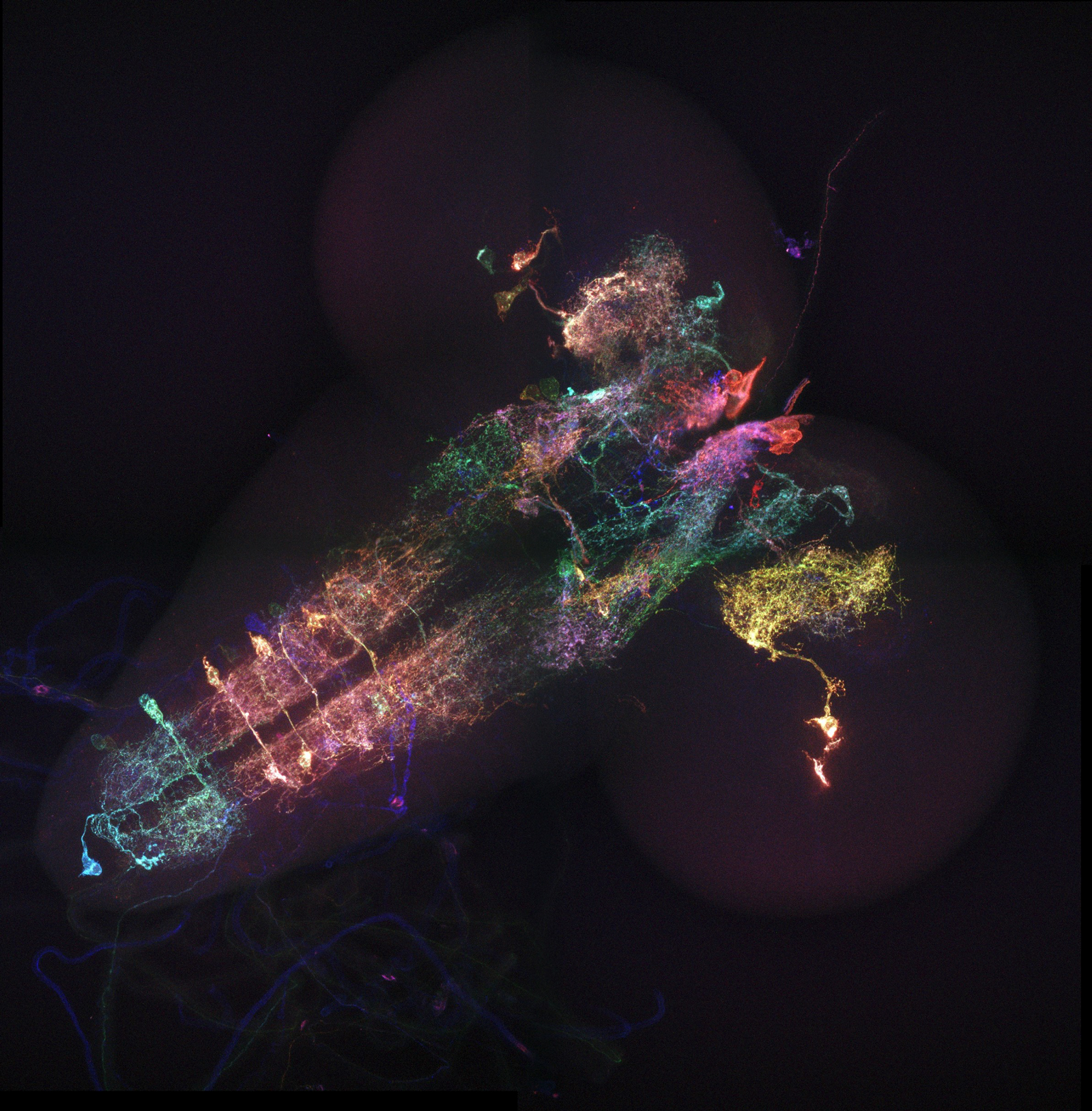
With the help of membrane-Bitbow (mBitbow), randomized fluorescent protein expression combinations are assigned to serotonergic neurons, revealing their patterned and diverse morphological features. The rich color palette provided by mBitbow makes serotonergic neurons’ complex neuropile densities well distinguished from each other, showing clear domains and branching patterns. mBitbow’s simple compact genetic design makes it readily compatible with the rich Gal4 driver libraries, providing a fast and powerful way to quickly assess morphology of neuron populations.
Image Credit: Ye Li
Li Y, Walker LA, Zhao Y, Edwards EM, Michki NS, Cheng HPJ, Ghazzi M, Chen TY, Chen M, Roossien DH, Cai D.
Bitbow Enables Highly Efficient Neuronal Lineage Tracing and Morphology Reconstruction in Single Drosophila Brains.
Front Neural Circuits. 2021 Oct 20;15:732183
Honorable Mention – Video
Bitbow Enables Highly Efficient Neuronal Lineage Tracing
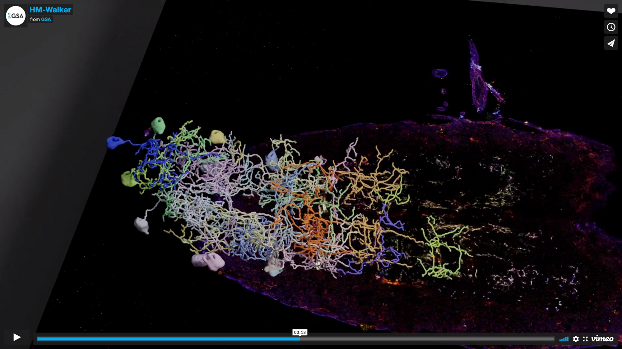
In this video, a 3D image stack of a larval Drosophila brain with Bitbow-labeled Serotonergic neurons is presented. Empowered by Bitbow’s high multi-color labeling capacity, and Protein-retention Expansion Microscopy (ProExM)’s physical resolution improvement, a complete tracing of 21 abdominal Serotonergic neurons (out of the estimated 26 total) is made possible from a single brain sample. 3D rendering of the tracing skeleton reveals distinct morphological groups of these neurons, highlighting the intrinsic heterogeneity within this critical population.
Video Credit: Logan Walker and Ye Li
i Y, Walker LA, Zhao Y, Edwards EM, Michki NS, Cheng HPJ, Ghazzi M, Chen TY, Chen M, Roossien DH, Cai D.
Bitbow Enables Highly Efficient Neuronal Lineage Tracing and Morphology Reconstruction in Single Drosophila Brains.
Front Neural Circuits. 2021 Oct 20;15:732183
Honorable Mention – Still Image
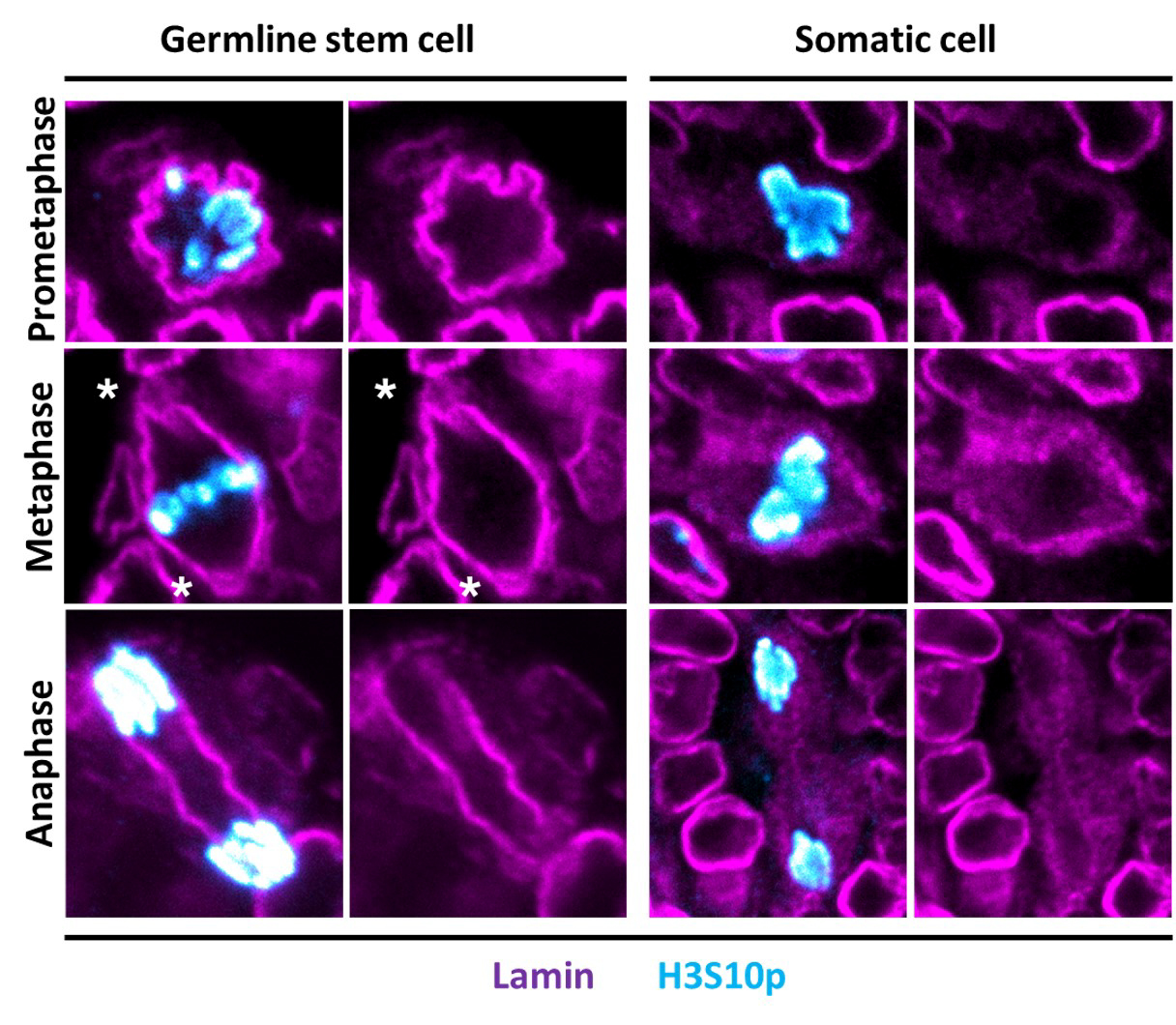
The nuclear envelope and nuclear lamina (NL) remain intact during mitosis of Germline Stem Cells (GSCs)
Show are representative images of mitotic GSCs and somatic cells in the adult ovary. These cells are stained with antibodies against a mitotic histone marker (Histone H3 Ser10 phosphorylation) and a marker of the NL (Lamin-B). Ovaries were also stained with antibodies against Vasa to identify germ cells (not shown). Stages of mitosis are shown to the left, based on the pattern of Histone H3 Ser10 phosphorylation. These data show that the GSC NL remains intact during mitosis, whereas the somatic cell NL disperses. Asterisks indicate positions of a remodeled NL in GSCs that holds the centrosome.
Image Credit: Tingting Duan
Duan T, Cupp R, Geyer PK.
Drosophila female germline stem cells undergo mitosis without nuclear breakdown.
Curr Biol. 2021 Apr 12;31(7):1450-1462
Honorable Mention – Video
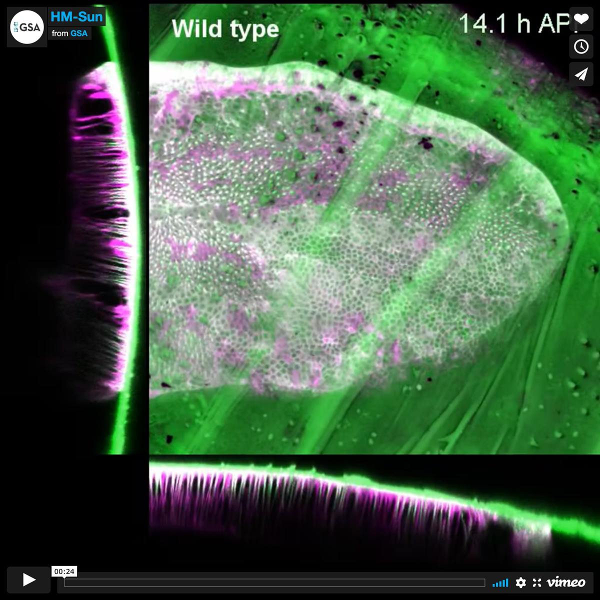
Microtubule-actin projections mediate Drosophila wing adhesion
Microtubule-actin projections mediate wing adhesionThe Drosophila wing is a flat appendage formed by mutual adhesion of its dorsal and ventral surfaces during metamorphosis. In the adhesion process, dorsal and ventral wing surfaces are kept in contact by long projections traversed by compact cytoskeletal cables combining F-actin (LifeAct-RFP, magenta) and microtubules (αTub-GFP, green). The video shows the final phase of the process (13 to 26 h after puparium formation), when microtubule-actin cables disassemble and projections quickly contract to bring the dorsal and ventral surfaces together. Knock down of extracellular matrix protein laminin (LanB2) prevents formation of projections. Projections form but do not contain compact cables upon knock down of spectraplakin (shot), septins (Sep1) or microtubule stabilizer Sdb. Confocal stacks were acquired through the pupal case every 3 min with a laser point scanning microscope. A whole-stack projection is shown next to XZ and YZ sections.
Video Credit: Tianhui Sun
Sun T, Song Y, Teng D, Chen Y, Dai J, Ma M, Zhang W, Pastor-Pareja JC.
Atypical laminin spots and pull-generated microtubule-actin projections mediate Drosophila wing adhesion.
Cell Rep. 2021 Sep 7;36(10):109667.
Honorable Mention – Still Image
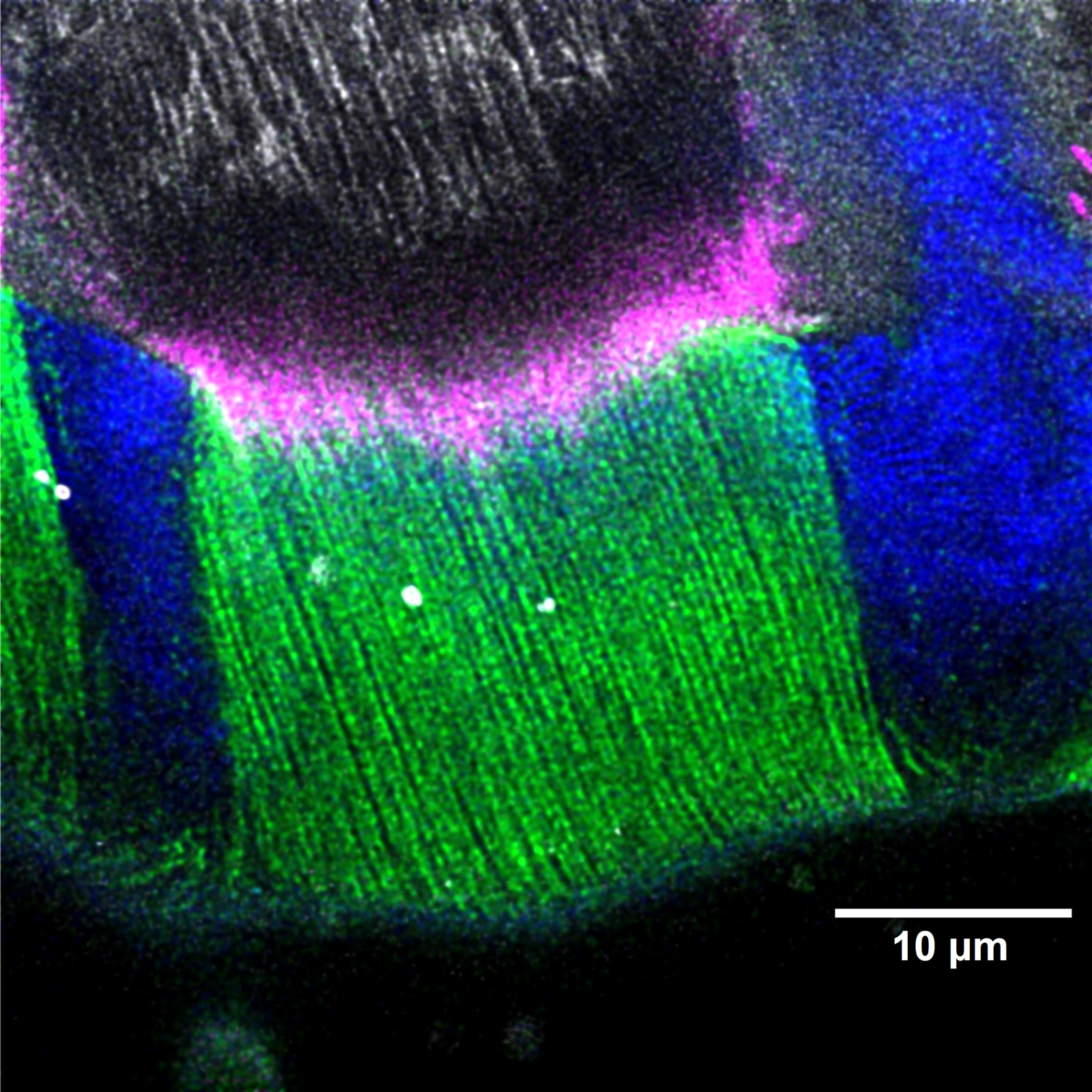
ECM nano-filaments in the tendon-exoskeleton attachment
To transmit the force generated by muscular contraction, the muscle-tendon system must be firmly attached to the exoskeleton of insects. Despite the muscle-tendon junction has been extensively studied, how tendon cells can anchor to the exoskeleton is much less understood. We found Drosophila Dumpy (Dpy), a giant apical ECM protein, forms nano-filaments that anchor the muscle-tendon system into the exoskeleton (cuticle) in the third instar larva. These nano-filaments are tightly bound to the microtubules in the tendon cells and the chitin in the exoskeleton. This figure shows the basic molecular anatomy of the tendon-exoskeleton attachments in the insect musculoskeletal system.
Image Credit: Wei-Chen Chu
Chu WC, Hayashi S.
Mechano-chemical enforcement of tendon apical ECM into nano-filaments during Drosophila flight muscle development.
Curr Biol. 2021 Apr 12;31(7):1366-1378
Honorable Mention – Video
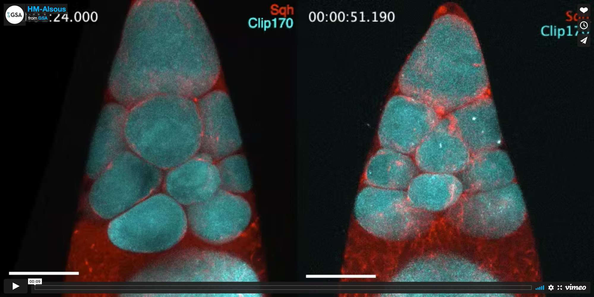
Actomyosin contractile waves cause cell deformations that facilitate cytoplasmic transport in Drosophila egg chambers
Actomyosin contractility is critical for numerous developmental processes. During the later stages of oocyte development in Drosophila melanogaster, fifteen sister ‘nurse cells’ connected to the oocyte rapidly transfer their contents through cytoplasmic bridges into the oocyte, providing it with the ingredients needed to support early embryonic development. Shown in the video are wave-like actomyosin contractions in the nurse cell clusters of two egg chambers, highlighting the resulting cell deformations that are required for cytoplasmic transport to run to completion. Red shows myosin regulatory light chain; cyan shows Clip170, a microtubule plus-end tracking protein. Scale bar: 50 µm.
Video Credit: Jasmin Imran Alsous
Imran Alsous J, Romeo N, Jackson JA, Mason FM, Dunkel J, Martin AC.
Dynamics of hydraulic and contractile wave-mediated fluid transport during Drosophila oogenesis.
Proc Natl Acad Sci U S A. 2021 Mar 9;118(10):e2019749118
