2021 Drosophila Image Award
Winner – Video
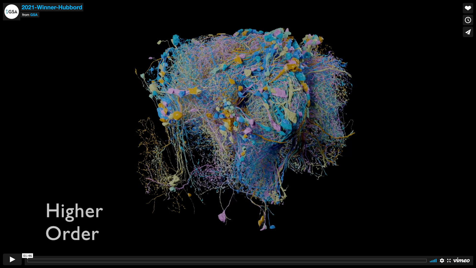
Janelia FlyEM Hemibrain Overview
The Janelia FlyEM hemibrain is the largest synaptic-level connectome ever reconstructed. It covers a large portion of the central Drosophila brain, including the mushroom body and central complex circuits critical for associative learning and fly navigation. This connectome reconstruction contains around 25,000 neurons, which can be grouped into thousands of distinct cell types spanning several brain regions; examples of the cell types for important regions appear in this video. Building this connectome required advances in imaging, segmentation (by the Connectomics Group at Google), and proofreading and analysis software. Analyzing this connectome is revealing new and unexpected insights into neural circuit structure, leading to improved theory and experimental design.
Video Credit: Philip Hubbard
Scheffer LK, …. , Plaza SM.
A connectome and analysis of the adult Drosophila central brain.
Elife. 2020 Sep 7;9:e57443.
Winner – Still Image
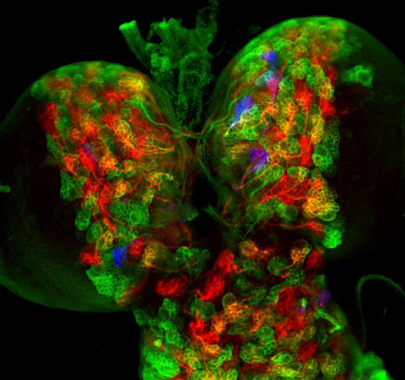
Drosophila larval brain labeled by targeting CLADES to progenitor cells
CLADES allows labeling cell lineages with a programmable sequence of fluorescent reporters. Drosophila larval brain labeled by targeting CLADES to progenitor cells (neuroblasts). Coupled to neuroblast division, CLADES labels progenitors with a cascade of colors that progresses throughout development. Neurons generated from these progenitors inherit a sequential cascade of reporters in order: green, yellow, red, purple and blue. Early- and late-born neurons become labeled with the first or the last colors in the sequence respectively
Image Credit: Jorge Garcia-Marques
Garcia-Marques, J., Espinosa-Medina, I., Ku, K., Yang, C., Koyama, M., Yu, H., & Lee, T.
(2020). A programmable sequence of reporters for lineage analysis.
Nature Neuroscience 2020 Dec;23(12):1618-1628
1st Runner Up
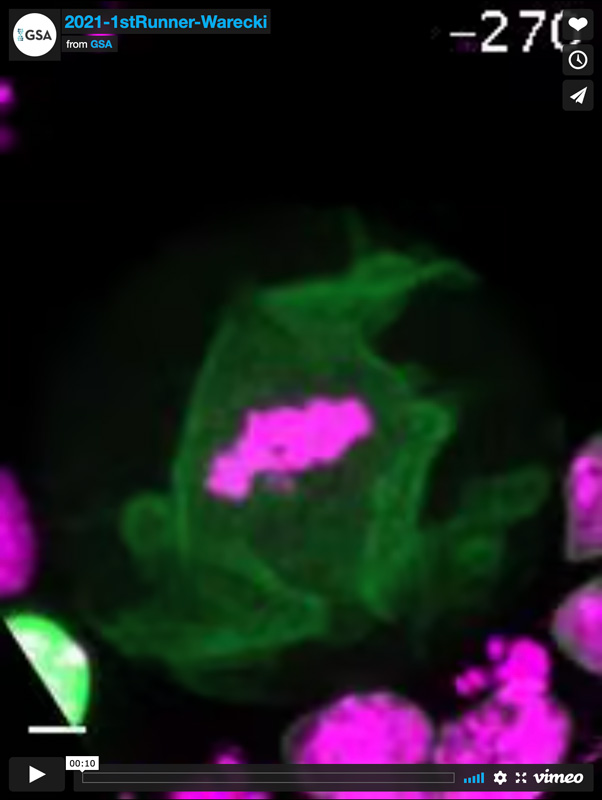
Lamin extensions guide broken chromosome fragments into telophase nuclei
Cells dividing with broken chromosome fragments must incorporate those fragments into daughter nuclei to maintain euploidy. However, mechanisms allowing incorporation of fragments into daughter nuclei remain poorly defined. This movie shows a neuroblast dividing with two chromosome fragments. Lamin extends from the nascent daughter nuclei towards to chromosome fragments, both shortening the distance the fragments must travel to join the daughter nuclei and also creating channels in the nascent nuclear envelopes surrounding daughter nuclei. The chromosomes fragments then pass through these channels to rejoin daughter nuclei and restore euploidy. Lamin is shown in green (elav-Gal4; UAS-Lam-GFP) and chromosomes in magenta (H2Av-RFP).
Video Credit: Brandt Warecki
Brandt Warecki, Ian Bast, Xi Ling, and William Sullivan.
ESCRT-III-mediated membrane fusion drives chromosome fragments through nuclear envelope channels.
Journal of Cell Biology 219, e201905091
1st Runner Up
EM reconstruction of a head direction system
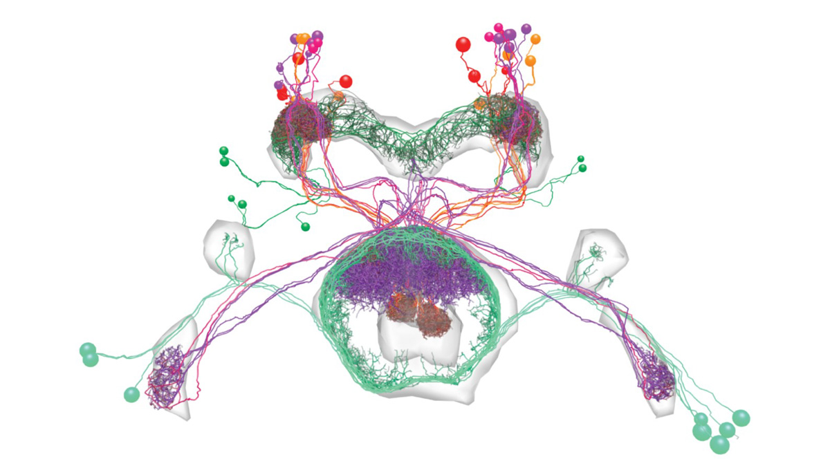
A head direction system in the Drosophila central complex is composed of multiple different cell types. Individual neurons from these cell types were reconstructed in an electron microscopy (EM) volume of the full adult fly brain (Zheng, Lauritzen et al., 2018). The connectivity between the reconstructed neurons is consistent with theoretical predictions for head direction networks. The figure shows the skeletons of the reconstructed neurons. Each cell type is rendered in a different color. The light gray regions show the outlines of neuropils.
Image Credit: Daniel Turner-Evans
Daniel B. Turner-Evans, Kristopher T. Jensen, Saba Ali, Tyler Paterson, Arlo Sheridan, Robert P. Ray, Tanya Wolff, Scott Lauritzen, Gerald M. Rubin, Davi Bock, Vivek Jayaraman
The neuroanatomical ultrastructure and function of a biological ring attractor.
Neuron 108, 145-163 (2020)
2nd Runner Up
Synapses between the dendrites of Kenyon cells and the olfactory projection neurons
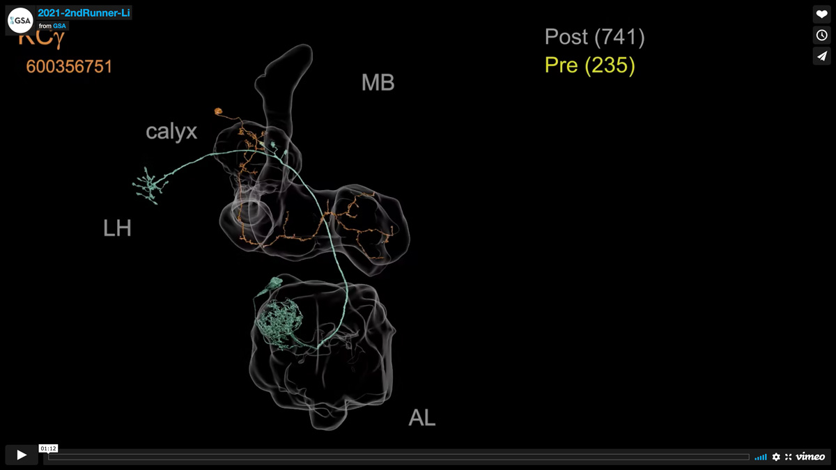
This narrated video illustrates the distinct structure of the synapses formed between the dendrites of Mushroom body Kenyon cells and the olfactory projection neurons that provide them with sensory input.
Video Credit: Feng Li (Video produced by Philip Hubbard)
Li F, Lindsey JW, Marin EC, Otto N, Dreher M, Dempsey G, Stark I, Bates AS, Pleijzier MW, Schlegel P, Nern A, Takemura SY, Eckstein N, Yang T, Francis A, Braun A, Parekh R, Costa M, Scheffer LK, Aso Y, Jefferis GS, Abbott LF, Litwin-Kumar A, Waddell S, Rubin GM.
The connectome of the adult Drosophila mushroom body provides insights into function.
Elife. 2020 Dec 14;9:e62576.
2nd Runner Up
Neurons that ‘talk’ to the gut
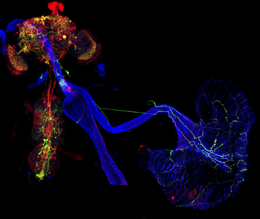
Neurons that ‘talk’ to the gut change their function to allow for the increase in food intake required for reproductive needs in females. A restricted subset of gut-innervating neurons that express the neuropeptide myosuppressin (Ms) innervate the crop, a stomach-like structure of the fly intestine. This figure shows co-expression between the Ms protein reporter (green) and Ms peptide (red) in the nervous system, and in neuronal projections towards the gut (blue).
Image Credit: Dafni Hadjieconomou
Dafni Hadjieconomou, George King, Pedro Gaspar, Alessandro Mineo, Laura Blackie, Tomotsune Ameku, Chris Studd, Alex de Mendoza, Fengqiu Diao, Benjamin H. White, André E. X. Brown, Pierre-Yves Plaçais, Thomas Préat & Irene Miguel-Aliaga.
Enteric neurons increase maternal food intake during reproduction.
Nature 587, 455–459 (2020)
Honorable Mention
Hindlegs are the key for backward walking
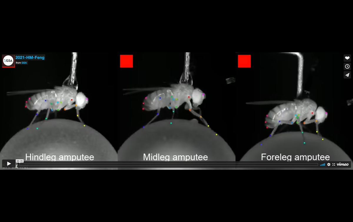
Moonwalker Descending Neurons (MDNs) are command type descending neurons that are necessary and sufficient for backward walking in Drosophila. Although innervating all three thoracic segments that each controls a pair of legs, MDNs impact hindlegs much more than others. Tarsal amputation experiments to remove ground grabbing power for each leg show only hindlegs are essential for propelling the body backwards when MDN is activated. Precise neuronal pathways executing MDN’s commands in metathoracic ganglion which controls hindleg movements have been identified in this study
Video Credit: Kai Feng
Feng K, Sen R, Minegishi R, Dübbert M, Bockemühl T, Büschges A, Dickson BJ.
Distributed control of motor circuits for backward walking in Drosophila.
Nat Commun. 2020 Dec 2;11(1):6166.
Honorable Mention
A gradient of ush mRNAs in response to graded BMP signaling in the early embryo
Graded Bone Morphogenic Protein (BMP) signaling patterns the dorsal ectoderm of the developing embryo and is interpreted by cells through modulation of transcriptional burst frequency. Different burst frequencies result in a gradient of mRNA numbers per cell shown in this figure for the BMP target gene ush. A fixed Drosophila embryo was stained with single molecule FISH probes targeting exonic ush regions (grey) and DAPI (blue). The number of cytoplasmic mRNA molecules was quantified per cell and is presented by false coloring the expression domain on the embryo and as a graph for the boxed region.
Image Credit: Caroline Hoppe
Caroline Hoppe, Jonathan R. Bowles, Thomas G. Minchington, Catherine Sutcliffe, Priyanka Upadhyai, Magnus Rattray and Hilary L. Ashe.
Modulation of the Promoter Activation Rate Dictates the Transcriptional Response to Graded BMP Signaling Levels in the Drosophila Embryo.
Dev Cell. 54, 727-741 (2020)
Honorable Mention
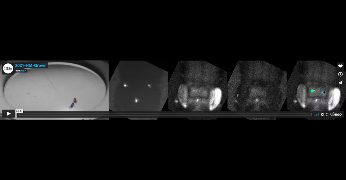
Activity of a cluster of ~20 P1 neurons during naturally evoked male courtship in Drosophila
Activity of a cluster of ~20 male-specific dorsal posterior fruitless gene-expressing P1 neurons during naturally evoked male courtship in Drosophila. A virgin male fly with P1a-Gal4, UAS-GCaMP6s, and UAS-myr-tdTomato transgenes (with the surgically created optically clear in vivo brain imaging window) being tracked in real-time (at 1,000 fps) while freely behaving and courting a female fly. Observed is the entire interaction sequence with increased activity in these neurons as the male courts the female fly. From left to right; arena-view, fly-view, fluo-view tdTomato channel, fluo-view GCaMP6s channel, and fluo-view tdTomato channel with a pseudo-color Fratio = FGCaMP6s/FtdTomato representation of the GCaMP6s signal in P1 neurons. In arena-view, blue dot represents tracked centroid of the male fly, and red dot represents tracked centroid of the female fly. Frames marked with the slashed-o symbol were excluded from fluorescence quantification based on image quality. Frame rate of video is reduced to 1/2x speed (25 Hz).
Video Credit: Dhruv Grover
Grover D, Katsuki T, Li J, Dawkins TJ, Greenspan RJ.
Imaging brain activity during complex social behaviors in Drosophila with Flyception2.
Nat Commun. 2020 Jan 30;11(1):623.
Honorable Mention
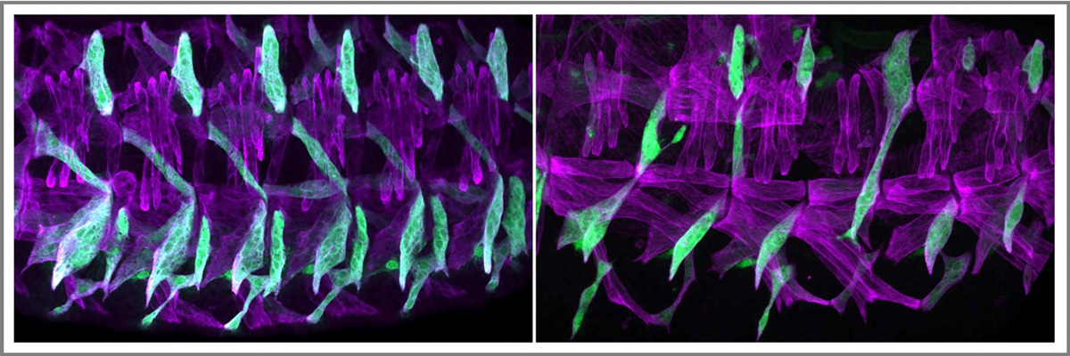
Morphological transformation of nascent myotubes
Nascent myotubes undergo a dramatic morphological transformation during myogenesis, in which the myotubes elongate over several cell diameters and are directed to the correct muscle attachment sites. Fibroblast Growth Factor (FGF) signaling receptor Heartless is enriched in nascent myotubes. This image shows the wild type (left panel) and htl mutant musculoskeletal system (right panels). htl mutant embryo showed significant myotube guidance defects. This result suggests that FGF signaling is essential for musculoskeletal system patterning during myogenesis.
Image Credit: Shuo Yang
Yang S, Weske A, Du Y, Valera JM, Jones KL, Johnson AN.
FGF signaling directs myotube guidance by regulating Rac activity.
Development. 2020 Feb 7;147(3)
Honorable Mention
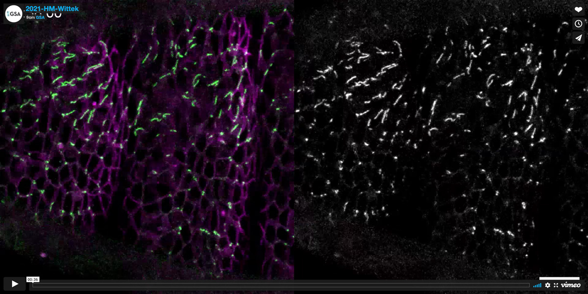
M6 is a tricellular occluding junction protein
M6 is a tricellular occluding junction protein that accumulates at epithelial cell vertices together with Anakonda and Gliotactin to form a tight paracellular barrier. The video shows a maximum intensity projection of lateral epidermis in a Dlg::mTagRFP;; GFP::M6 embryo (stage 13). Anterior is to the left, dorsal is up. GFP::M6 (green in merge) is initially distributed around lateral membranes and begins to accumulate at vertices in segmental patches in the dorsal epidermis. Note that GFP::M6 accumulation at vertices spreads from dorsal to ventral in the epidermis, and that GFP::M6 signals extend in an apical-to-basal direction along each vertex. This can be seen in the depth-coded time series below the movie. Images were acquired at 30s intervals. Time is indicated (hh:mm:ss).
Video Credit: Anna Wittek
Wittek A, Hollmann M, Schleutker R, Luschnig S.
The Transmembrane Proteins M6 and Anakonda Cooperate to Initiate Tricellular Junction Assembly in Epithelia of Drosophila.
Curr Biol. 2020 Nov 2;30(21):4254-4262
Honorable Mention
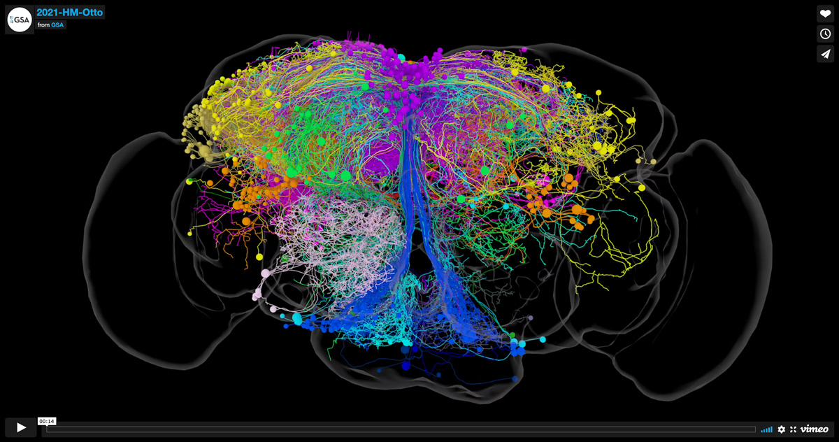
Neurons providing input to dopaminergic neurons of the mushroom body
Nanoscale 3D representation of neurons providing input to dopaminergic neurons that innervate the adult fly mushroom body. Different types of dopaminergic neurons reinforce memories of opposite valence, and some provide state-dependent motivational control. The image shows a selection of their input neurons that were manually reconstructed from the EM-image volume of a full adult female brain (FAFB) and coarsely clustered by morphology. These morphological groupings are shown in different colors.
Video Credit: Nils Otto
Otto N, Pleijzier MW, Morgan IC, Edmondson-Stait AJ, Heinz KJ, Stark I, Dempsey G, Ito M, Kapoor I, Hsu J, Schlegel PM, Bates AS, Feng L, Costa M, Ito K, Bock DD, Rubin GM, Jefferis GSXE, Waddell S.
Input Connectivity Reveals Additional Heterogeneity of Dopaminergic Reinforcement in Drosophila.
Curr Biol. 2020 Aug 17;30(16):3200-3211
