2017 Drosophila Image Award
Winner – Video
Systems Analysis of the Dynamic Inflammatory Response to Tissue Damage
Following tissue damage, a complex repair process is initiated to restore the integrity of the injured tissue. Re-epithelialisation is accompanied by a rapid inflammatory response, whereby cells of the innate immune system (here, Drosophila macrophages called ‘hemocytes’) are rapidly recruited to the damaged area. Although we can live-image this process with high spatial and temporal resolution in vivo, the precise identity and properties of the immune damage attractants remain unclear.
Here, we employ an alternative computational approach to extract detailed information about the wound attractant signal from the in vivo dynamic behaviour of the responding immune cells. Using this novel approach, we have uncovered new insight into the spatio-temporal properties of the wound attractant, revealing precisely how the damage signal spreads out from the injury site in space and time, and inferring parameters such as the diffusion coefficient, source, and duration of active signal production. Our study highlights the valuable insight that can be extracted from in vivo imaging data if more sophisticated analysis tools are employed, that would otherwise remain experimentally inaccessible.
Weavers H, Liepe J, Sim A, Wood W, Martin P & Stumpf M. (2016)
Systems Analysis of the Dynamic Inflammatory Response to Tissue Damage Reveals Spatiotemporal Properties of the Wound Attractant Gradient
Current Biology 26, 1975-1989 (2016)
doi:10.1016/j.cub.2016.06.012
Winner – Still Image
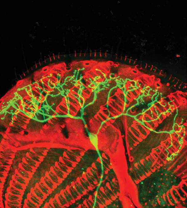
The basis of food texture sensation in Drosophila
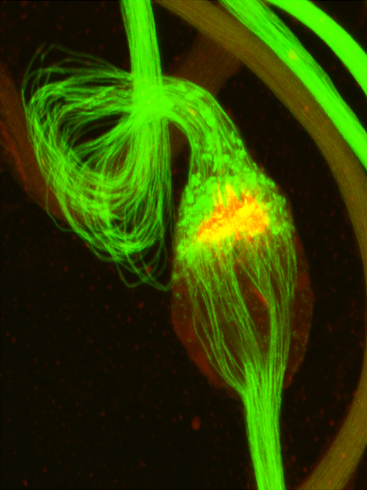
Food texture such as the degree of hardness, plays a very important role in regulating food preference in animals. In the fruit fly, food texture sensation depends on a multidendritic neuron (md-L) in its tongue – the labellum. The mechanical forces produced by food chewing activate the md-L neuron, which in turn transmits food texture information to the brain. Shown is an md-L neuron (green) extending elaborate dendrites to the bases of many taste hairs. The red shows rows of spring-like structures in the fly tongue.
Reference: Zhang, Y.V., Aikin, T.J., Li, Z., and Montell, C. (2016)
The basis of food texture sensation in Drosophila.
Neuron 91, 863-877. PMID: 27478019
Non-invasive Tension Estimation during the Early Stage of Dorsal Closure
Time-lapse movie of an Drosophila embryo expressing Dα-cateninRFP (left), the estimated tension (middle), and the hotspot analysis (right).
We used amnioserosa cells, which exhibit rapid cell oscillation during Drosophila dorsal closure, and reported that cell junctional tension closely correlated with boundary dynamics and shapes, i.e., the contracting and straighter boundary has more tension, while elongating and wavy boundary has less tension. Based on such correlations, we non-invasively estimated a tension map across the tissue, and found that the tension dynamically fluctuates during boundary oscillation. Moreover, this estimations helped us to find that an adherens junction molecule, vinculin, dynamically accumulates to or dissociates from oscillating boundary in a junctional-tension-dependent manner. This study showed the basic mechanics of cell boundary deformations, their spatiotemporal evolution on a tissue, and accompanying molecular dynamics.
Yusuke Hara, Murat Shagirov, and Yusuke Toyama
Cell boundary elongation by non-autonomous contractility in cell oscillation.
Current Biology 26, pp2388–2396 (2016)
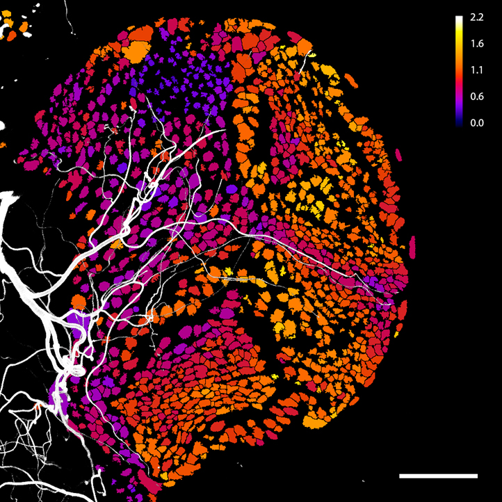
Different cellular hypoxic states correlate with tracheal supply in the brain.
The image shows a confocal section of a brain hemisphere of a Drosophila third-instar larva. Nuclei were segmented and assigned a colour according to the readout of a ratiometric hypoxia biosensor. Blue corresponds to highest oxygenation, while yellow corresponds to lowest oxygenation. The image is superimposed by a maximum intensity projection of the brain’s tracheal system (white). Note the topographical correlation between the extent of tracheal supply and the distinct hypoxic states of different brain regions.
Tvisha Misra, Martin Baccino-Calace, Felix Meyenhofer, David Rodriguez-Crespo, Hatice Akarsu, Ricardo Armenta-Calderón, Thomas A. Gorr, Christian Frei, Rafael Cantera, Boris Egger and Stefan Luschnig
A genetically encoded biosensor for visualizing hypoxia responses in vivo
Biology Open (2016). doi:10.1242/bio.018226
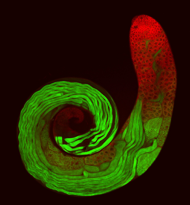
Spermatogenesis in the Drosophila melanogaster testis
Spermatogenesis is an ongoing developmental process that requires the continual production of new germ cells from an adult stem cell niche. In Drosophila, the stem cell niche is made up of a cluster of somatic cells called the hub located at the apex of the testis (top of this image). In order for germ cells to differentiate, they must be encapsulated by somatic cyst cells, which isolate the germ cells by forming occluding septate junctions. If septate junctions are disrupted the germline fails to differentiate while the somatic cyst cells switch fate and form new hub cells. The larger hub recruits more stem cells, increasing the size of the niche. This suggests that the stable size of the stem cell niche in adult testes is actively maintained by signals from the differentiating cells outside of the niche itself. This image shows germline differentiation during spermatogenesis with pre-meiotic germ cells labelled by Vasa (red), and post-meiotic germ cells labelled by β2-tubulin (green).
Fairchild, M.J., Yang, L., Goodwin, K., Tanentzapf, G.
Occluding junctions maintain stem cell niche homeostasis in the fly testis.
Current Biology, 26(18):2492–2499 (2016).
Variation among Drosophila species in investment per sperm and in spermatogenesis.
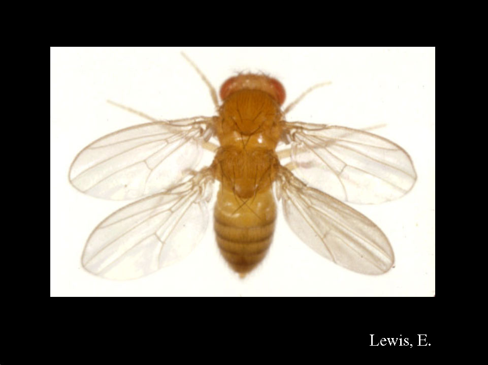
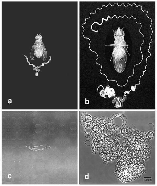
The Drosophila lineage has experienced greater diversification in sperm length than the remainder of the animal kingdom, in part due to female-mediated post-copulatory sexual selection. Longer sperm are energetically costlier to manufacture. As part of a study to discern the adaptive value of the female post-copulatory preference for long sperm, a comparative investigation examined the sex-specific allometry of reproductive potential using seven Drosophila species. This figure shows an intact male fly above his reproductive tract (a, b) and a single spermatozoon (c, d) for D. arizonae (a, c) and D. bifurca (b, d), which produce the shortest and longest sperm, respectively, in the study. The 58.3 mm long sperm of D. bifurca are 38 times longer than those of D. arizonae and, despite making very few sperm, D. bifurca males make an over three-fold greater investment in spermatogenesis than do D. arizonae males. In phylogenetic regressions, the male reproductive potential became increasingly condition-dependent as sperm length increased. Hence, males of any condition can produce and inseminate many “cheap” sperm, but only high-quality males have the available resources to produce abundant “expensive” sperm. All panels depict equal magnification.
Lüpold, S., M. K. Manier, N. Puniamoorthy, C. Schoff, W. T. Starmer, S. H. Buckley Luepold, J. M. Belote and S. Pitnick. 2016.
How sexual selection can drive the evolution of costly sperm ornamentation. Nature 533: 535-538.
A Krebs Cycle Component Limits Caspase Activation Rate through Mitochondrial Surface Restriction of CRL Activation.
The image shows terminally differentiating Drosophila spermatids during the caspase-dependent vital cellular process of “individualization”. During this process, spermatids extrudes their bulk cytoplasmic contents into a “waste bag” (marked by the “individualization complex”; yellow/orange), yielding highly compact mature sperm cells. The Krebs cycle component, A-Sβ (green), is expressed on the surface (rather than the matrix) of the mitochondria, where it binds to and activates a Ub ligase complex required for caspase activation. This new function of A-Sβ limits the rate of caspase activation by restricting the active Ub ligase complex to the vicinity of the mitochondria.
Lior Aram, Tslil Braun, Carmel Braverman, Yosef Kaplan, Liat Ravid, Smadar Levin-Zaidman and Eli Arama.
DEVELOPMENTAL CELL.37:15-33 (2016).
Defensive kicking against invading mites
Parasitic mites present threats to many insect species. We used a high-speed camera to record how fruit flies responses to invading mites. As shown in the movie (playback speed slowed down to 1/100x), when a mite touched the wing margin of a fly, it kicked the mite off with a stereotyped behavioral sequence: sensing the presence of the moving mite immediately after the mite reached its wing margin, pulling back the body, swinging the hind leg on the ipsilateral side and then kicking the mite off. The entire behavioral sequence was completed within 200 milliseconds with high spatial precision — fast and accurate enough to kick off a moving mite. Our further study identified the mechanosensory machinery underlying this defensive kicking behavior, and revealed that the spatial precision only requires VNC circuits but not the brain.
Jiefu Li, Wei Zhang, Zhenhao Guo, Sophia Wu, Lily Yeh Jan and Yuh-Nung Jan.
A Defensive Kicking Behavior in Response to Mechanical Stimuli Mediated by Drosophila Wing Margin Bristles.
Journal of Neuroscience 2 November 2016, 36 (44) 11275-11282.
A Taste Circuit that Regulates Ingestion by Integrating Food and Hunger Signals
Real-time GCaMP6s imaging of IN1 cholinergic interneuron arbors in the brain of a 24 hour fasted female fly offered a 20 nl drop of 1 M sucrose. The green square marks the period of ingestion.
Yapici, N., Cohn, R., Schusterreiter, C., Ruta, V., & Vosshall, L. B. (2016).
A taste circuit that regulates ingestion by integrating food and hunger signals.
Cell, 165(3), 715-729.
