2016 Drosophila Image Award
Winner – Still Image
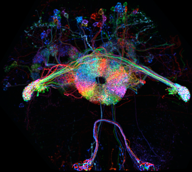
“Burning Man” Showcases Multicolor Flip-out Technique in Drosophila Brain
Neurons labeled using the multicolor flip-out strategy, which stochastically labels small subsets of cells in distinct colors, reveal the organization of the integration center of the Drosophila brain, the central complex, at a single cell level. This technique enabled the identification and detailed morphological characterization of a subset of neurons that arborize throughout this key integration center. This analysis also provided a set of GAL4 lines that identifies subsets of neurons in the neuropils that constitute the central complex. Two neuropils of the central complex are evident in this image, the protocerebral bridge and donut-shaped ellipsoid body.
Tanya Wolff, Nirmala A. Iyer and Gerald M. Rubin.
Neuroarchitecture and neuroanatomy of the Drosophila central complex: A GAL4-based dissection of protocerebral bridge neurons and circuits.
Journal of Comparative Neurology 523, 997-1037 (2015).
Winner – Video
IsoView whole-animal functional imaging of larval Drosophila
Lateral (left), dorsoventral (middle) and rotating (right) maximum-intensity projections of a multi-view deconvolved time-lapse recording of an early Drosophila first instar larva expressing the calcium indicator GCaMP6s throughout the nervous system. The data set was acquired with a custom-built light-sheet microscope for isotropic multi-view imaging (IsoView microscope), which provides isotropic, sub-cellular spatial resolution in all three dimensions. Functional imaging was performed at 2 Hz. The images are gamma-corrected and shown using a false-color look-up-table (blue to yellow) to reduce the high dynamic range of the raw data for better visibility.
Raghav Chhetri, Fernando Amat, Yinan Wan, Burkhard Höckendorf, William Lemon and Philipp Keller.
Whole-animal functional and developmental imaging with isotropic spatial resolution.
Nature Methods 12, 1171-8 (2015).
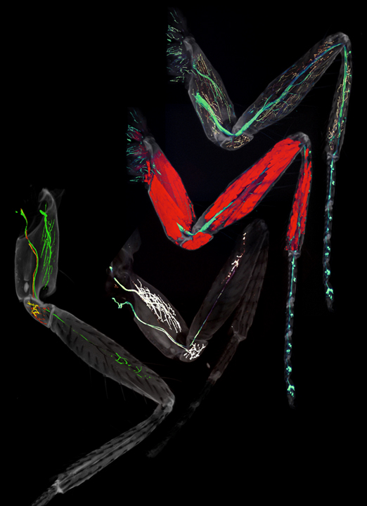
Visualization of the leg axonal targeting: from 50 Motoneurons to single Motoneurons
From top right to bottom left: 1) T1 leg expressing mCD8::GFP (blue/green, depending on intensity) and the synaptic marker rab3::YFP (yellow) under the control of VGlut-Gal4. 2) T1 leg expressing mCD8::GFP (blue/green, depending on intensity) under the control of VGlut-Gal4 and Mhc (Myosin heavy chain)-RFP. 3) T1 leg containing a neuroblast lineage MARCM clone called lineage B (Lin B) and expressing mCD8::GFP (blue/green, depending on intensity) and the synaptic marker rab3::YFP (yellow) under the control of VGlut-Gal4. 4) T1 leg containing a Lin B MARCMbow clone where all LinB motor neurons express mCD8::GFP (green). Two motor neurons called Tr1 and Tr2 were independently labeled with CD8::mCherry (red) and pm-mCitrine (yellow), respectively. These techniques have allowed us to visualize all motor neurons, specific neuroblast lineages and finally single motor neurons in order to analyze phenotypes at the single level and to demonstrate that the individual morphology of motor neurons is dictated by a combinatorial code of transcription factors.
Enriquez J, Venkatasubramanian L, Baek M, Peterson M, Venkatasubramanian L, Aghayeva U and Mann RS.
Specification of individual adult motor neuron morphologies by combinatorial transcription factor codes. Neuron. 2015 May 20;86(4):955-70. Co-corresponding author, PMCID: PMC4441546
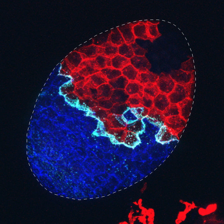
Visualizing cellular contacts at clonal boundaries
Genetic mosaics are a powerful tool to analyze cell-cell interactions within a tissue because the behavior of lineage-related groups of cells (clones) can be tracked by cellular markers. We developed a new system to generate genetic mosaics called “CoinFLP”, which enables independent control of gene expression in two clonal populations. This feature of CoinFLP allows expression of two halves of a membrane-targeted split-GFP protein in two separate clonal populations. Therefore, cellular contacts between the two clonal populations will reconstitute fluorescent GFP (“GRASP”), allowing pronounced visualization of clonal boundaries. Here, we show a confocal projection of an egg chamber (outlined) composed of follicle cells that express LexA (red), Gal4 (blue), or neither (black). GRASP (cyan) labels the contacts between LexA-expressing cells and Gal4-expressing cells. Thus, the CoinFLP system enables visualization of each clonal population and their cellular contacts in one image.
Bosch, J. A., Tran, N. H., & Hariharan, I. K.
(2015). CoinFLP: a system for efficient mosaic screening and for visualizing clonal boundaries in Drosophila. Development, 142(3), 597–606.
IsoView multi-color imaging of Drosophila gastrulation
From left to right: dorsal, ventral, lateral-left, lateral-right and rotating maximum-intensity projections of a multi-view deconvolved time-lapse recording of a Drosophila embryo ubiquitously expressing GFP targeted to membranes and RFP targeted to cell nuclei (colors are inverted for better visibility). The data set was acquired with a custom-built light-sheet microscope for isotropic multi-view imaging (IsoView microscope), which provides isotropic, sub-cellular spatial resolution in all three dimensions. The video shows embryonic development during stages 6-8. Image data were acquired at 4-second intervals in and are displayed using gamma correction to reduce the high dynamic range of the raw data for better visibility.
Raghav Chhetri, Fernando Amat, Yinan Wan, Burkhard Höckendorf, William Lemon and Philipp Keller.
Whole-animal functional and developmental imaging with isotropic spatial resolution.
Nature Methods 12, 1171-8 (2015).
Transcriptional Memory in the Drosophila Embryo
By monitoring the expression of stochastically expressed transgenes in living Drosophila embryos, we visualize and quantify the extent to which transcription is inherited from mother to daughter cells in a living embryo. MS2 nascent mRNAs are shown in green (MS2-MCP-GFP) and nuclei are labeled in red (Histone-mRFP).
Here we show a false-colored snaE<sogPr<MS2 transgenic embryo, imaged from cc13 to gastrulation, the first 23 minutes of cc14 are shown. All active nuclei at cc13 are tracked with a unique number. After mitosis their descendants are tracked, circled in yellow, while active cc14 nuclei derived from inactive mothers are circled in blue.
Teresa Ferraro, Emilia Esposito, Laure Mancini, Sam Ng, Tanguy Lucas, Mathieu Coppey, Nathalie Dostatni, Aleksandra M. Walczak, Michael Levine and Mounia Lagha.
Transcriptional memory in the Drosophila embryo. Curr Biol. 2015 Dec 30. pii: S0960-9822(15)01506-7. doi: 10.1016/j.cub.2015.11.058
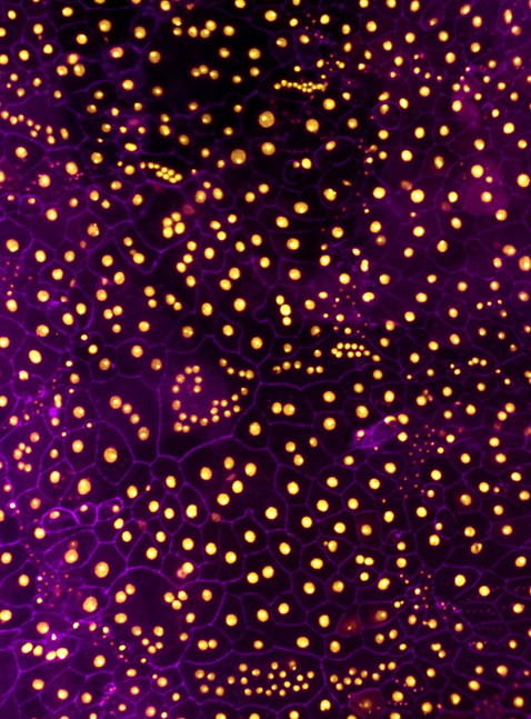
Loss of PINCH alters the stereotypical architecture of Drosophila larval epidermis
Drosophila larval epidermal cells are typically mononucleate and highly ordered, assuming polygonal shapes of a remarkably uniform size. The image shows a larval epidermal sheet deficient for the integrin adhesion adaptor protein PINCH. Epidermal membranes (anti-fasciclin III, magenta), and nuclei (expressing DsRed2Nuc, orange) are labeled. PINCH knockdown leads to variously sized and misshapen multinucleate cells—some containing in excess of 20 nuclei. The findings are important because the integrin complex, although previously implicated in many cellular functions in diverse tissues, has not previously been shown to be a suppressor of cell-cell fusion.
Yan Wang, Marco Antunes, Aimee E. Anderson, Julie L. Kadrmas, Antonio Jacinto, Michael J. Galko.
Integrin Adhesions Suppress Syncytium Formation in the Drosophila Larval Epidermis.
Curr Biol. 2015 Aug 31;25(17):2215-27. doi: 10.1016/j.cub.2015.07.031. Epub 2015 Aug 6.
Nanobody-mediated morphogen trapping
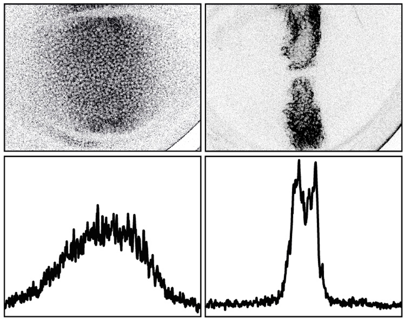
Morphogen gradient have been suggested to control patterning and growth of developing organs. To investigate the impact of morphogen spreading on patterning and growth control, we developed a nanobody-based method, we named morphotrap, that allows modification of morphogen gradient shape of GFP-tagged morphogens in vivo.
An EGFP-tagged version of Dpp forms a wide extracellular concentration gradient in the wing disc (left). Expression of morphotrap in Dpp producing cells results in retention of EGFP::Dpp along the surface of producing cells and results in a complete loss of the Dpp gradient (right). The concentration profiles are shown below the respective images.
Using morphotrap-mediated EGFP::Dpp trapping, we showed that Dpp spreading is required for patterning and growth control of the wing disc tissue, however is not required to control the proliferation rate of wing disc cells. Morphotrap offers a novel and versatile way to study the localization, dispersal and dynamics of morphogens and other secreted proteins.
Harmansa, S., Hamaratoglu, F., Affolter, M., Caussinus, E.
Dpp spreading is required for medial but not for lateral wing disc growth. Nature 527(7578):317-22. (2015) doi: 10.1038/nature15712
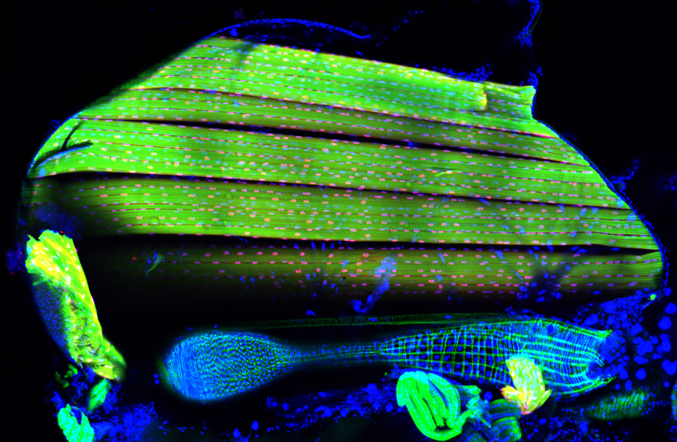
Flight muscles – Bruno regulates alternative splicing
Drosophila flight muscles are gigantic cells of 1mm length, containing hundreds of nuclei (red) and thousands of myofibrils (green) per muscle fiber. The particular contractile apparatus of these muscles enables very rapid muscle contractions and thus flight.
We showed here that the formation of this particular contractile apparatus requires alternative splicing of several hundred mRNAs, many coding for sarcomeric proteins. Alternative splicing in the flight muscle mode is regulated by the RNA binding protein Arrest/Bruno that is specifically expressed only during development of the flight muscles.
Dus, M et al. Nutrient Sensor in the Brain Directs the Action of the Brain-Gut Axis in Drosophila. 2015, Neuron 87, 1–13.
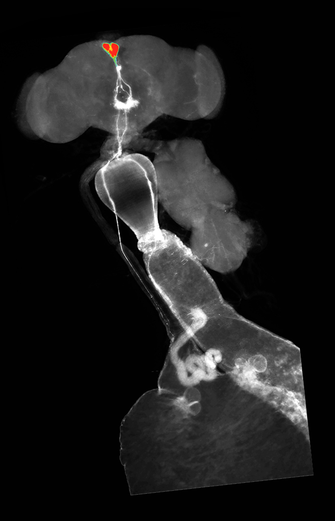
Sugar is both sweet and nutritive. While the taste cells that detect its sweetness were known, those that sense its nutritional value were not. This image shows the anatomy of the circuit that senses the nutritional value of sugar.
Cells expressing the fly neuropeptide Diuretic Hormone 44, the homologue of fly Corticotropin releasing factor, are located in the pars intercelebralis and send their projections to the crop and gut. The Dh44 cells respond exclusively to nutritive sugars, shown here a heatmap pseudocolor obtained from Calcium imaging experiments where fly brains were perfused with 20mM D-glucose. Nutrient perception in animals is thus mediated by a glucosensing brain-gut axis that reinforces the consumption of nutritive foods.
Stem cell nanotubes
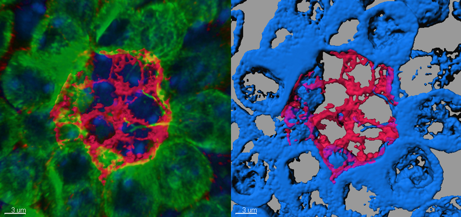
Male germline stem cells possess unique cellular protrusions, microtubule-based nanotubes (MT-nanotubes). MT-nanotubes are observed specifically within stem cell populations, and promote Dpp signal reception from the niche, contributing to the short-range nature of niche–stem-cell signalling.
Left: Three-dimensional images of a testicular niche in with Armadillo staining (Red: Hub cell coltex). nos-gal4>GFP-alpha-tubulin(green) illuminates MT-nanotubes which penetrate into neighboring niche (hub) cells (Armadillo; red). DAPI (blue).
Right: Surface rendering image of Armadillo (red) and GFP–alpha-tubulin (blue). Three-dimensional rendering was performed by Imaris software.
M. Inaba, M. Buszczak, and Y. M. Yamashita
“Nanotubes mediate niche-stem-cell signalling in the Drosophila testis.,” Nature, vol. 523, no. 7560, pp. 329–332, Jul. 2015.
Role of the SLC36 Pathetic in supporting extreme dendrite growth
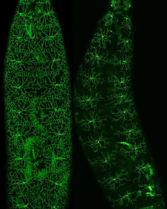
The SLC36 family member Pathetic is required for extreme dendrite growth in Drosophila somatosensory neurons. Shown here are confocal images of Drosophila Class IV da neurons (labeled by ppk-CD8-GFP) in wild type (left) or pathetic mutant 2nd instar larva (right). Although early growth and patterning is normal, dendrite growth arrests at a fixed limit in pathetic mutants. Mutation of pathetic selectively affects dendrite growth in neurons with large dendrite arbors, doing so by affecting translational output of neurons. However, pathetic is dispensable for dendrite growth in neurons with small dendrite arbors or the animal overall, demonstrating that growth of large dendrite arbors entails specialized molecular mechanisms.
Lin WY, Williams C, Yan C, Koledachkina T, Luedke K, Dalton J, Bloomsburg S, Morrison N, Duncan KE, Kim CC, Parrish JZ.
The SLC36 transporter Pathetic is required for extreme dendrite growth in Drosophila sensory neurons.
Genes Dev. 2015 Jun 1;29(11):1120-35. doi: 10.1101/gad.259119.115.
